| Reactivity: | Human |
| Applications: | WB, IF/ICC, ELISA |
| Host Species: | Rabbit |
| Isotype: | IgG |
| Clonality: | Polyclonal antibody |
| Gene Name: | catechol-O-methyltransferase |
| Gene Symbol: | COMT |
| Synonyms: | HEL-S-98n; [KD Validated] COMT |
| Gene ID: | 1312 |
| UniProt ID: | P21964 |
| Immunogen: | Recombinant fusion protein containing a sequence corresponding to amino acids 51-271 of human COMT. (NP_000745.1). |
| Dilution: | WB 1:500-1:2000 |
| Purification Method: | Affinity purification |
| Concentration: | 0.79 mg/mL |
| Buffer: | PBS with 0.05% proclin300, 50% glycerol, pH7.3. |
| Storage: | Store at -20°C. Avoid freeze / thaw cycles. |
| Documents: | Manual-COMT polyclonal antibody |
Background
Catechol-O-methyltransferase catalyzes the transfer of a methyl group from S-adenosylmethionine to catecholamines, including the neurotransmitters dopamine, epinephrine, and norepinephrine. This O-methylation results in one of the major degradative pathways of the catecholamine transmitters. In addition to its role in the metabolism of endogenous substances, COMT is important in the metabolism of catechol drugs used in the treatment of hypertension, asthma, and Parkinson disease. COMT is found in two forms in tissues, a soluble form (S-COMT) and a membrane-bound form (MB-COMT). The differences between S-COMT and MB-COMT reside within the N-termini. Several transcript variants are formed through the use of alternative translation initiation sites and promoters.
Images
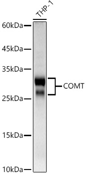
 | Western blot analysis of lysates from THP-1 cells using [KD Validated] COMT Rabbit pAb (A23600) at 1:700 dilution. Secondary antibody:HRP Goat Anti-Rabbit IgG (H+L)( AS014) at 1:10000 dilution. Lysates/proteins: 25 μg per lane. Blocking buffer: 3% nonfat dry milk in TBST. Detection: ECL Basic Kit (RM00020). Exposure time: 10s. |
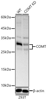
 | Western blot analysis of lysates from wild type (WT) and COMT knockdown (KD) 293T cells using [KD Validated] COMT Rabbit pAb (A23600) at 1:700 dilution. Secondary antibody: HRP-conjugated Goat anti-Rabbit IgG (H+L) (AS014) at 1:10000 dilution.Lysates/proteins: 25 μg per lane. Blocking buffer: 3% nonfat dry milk in TBST. Detection: ECL Basic Kit (RM00020). Exposure time: 10s. |
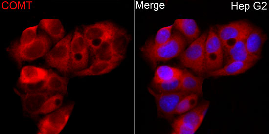
 | Immunofluorescence analysis of HepG2 cells using [KD Validated] COMT Rabbit pAb (A23600) at dilution of 1:50 (40x lens). Secondary antibody: Cy3-conjugated Goat anti-Rabbit IgG (H+L) (AS007) at 1:500 dilution. Blue: DAPI for nuclear staining. |
You may also be interested in:


