| Reactivity: | Human |
| Applications: | WB, IHC-P, IF/ICC, ELISA |
| Host Species: | Rabbit |
| Isotype: | IgG |
| Clonality: | Polyclonal antibody |
| Gene Name: | Epidermal growth factor receptor |
| Gene Symbol: | EGFR |
| Synonyms: | ERBB; ERRP; HER1; mENA; ERBB1; PIG61; NISBD2; FR |
| Gene ID: | 1956 |
| UniProt ID: | P00533 |
| Immunogen: | Recombinant fusion protein containing a sequence corresponding to amino acids 1021-1210 of human EGFR (NP_005219.2). |
| Dilution: | WB 1:500-1:1000 |
| Purification Method: | Affinity purification |
| Concentration: | 2.49 mg/ml |
| Buffer: | PBS with 0.05% proclin300, 50% glycerol, pH7.3. |
| Storage: | Store at -20°C. Avoid freeze / thaw cycles. |
| Documents: | Manual-EGFR polyclonal antibody |
Background
The protein encoded by this gene is a transmembrane glycoprotein that is a member of the protein kinase superfamily. This protein is a receptor for members of the epidermal growth factor family. EGFR is a cell surface protein that binds to epidermal growth factor, thus inducing receptor dimerization and tyrosine autophosphorylation leading to cell proliferation. Mutations in this gene are associated with lung cancer. EGFR is a component of the cytokine storm which contributes to a severe form of Coronavirus Disease 2019 (COVID-19) resulting from infection with severe acute respiratory syndrome coronavirus-2 (SARS-CoV-2).
Images
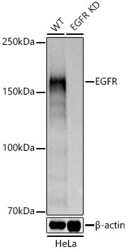 | Western blot analysis of lysates from wild type(WT) and EGFR knockdown (KD) HeLa cells, using [KD Validated] EGFR Rabbit pAb (A11577) at 1:1000 dilution. Secondary antibody: HRP-conjugated Goat anti-Rabbit IgG (H+L) (AS014) at 1:10000 dilution. Lysates/proteins: 25μg per lane. Blocking buffer: 3% nonfat dry milk in TBST. Detection: ECL Basic Kit (RM00020). Exposure time: 0.5s. |
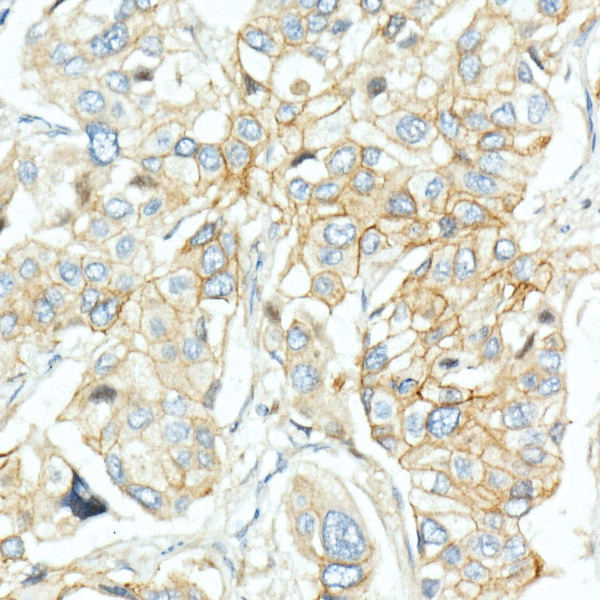 | Immunohistochemistry analysis of paraffin-embedded Human lung cancer using [KD Validated] EGFR Rabbit pAb (A11577) at dilution of 1:20 (40x lens). High pressure antigen retrieval performed with 0.01M Citrate Bufferr (pH 6.0) prior to IHC staining. |
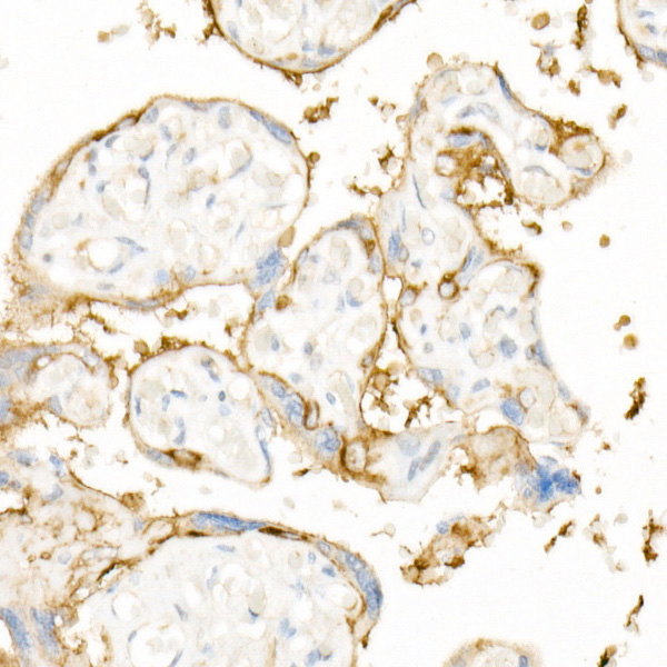 | Immunohistochemistry analysis of paraffin-embedded Human placenta using [KD Validated] EGFR Rabbit pAb (A11577) at dilution of 1:20 (40x lens). High pressure antigen retrieval performed with 0.01M Citrate Bufferr (pH 6.0) prior to IHC staining. |
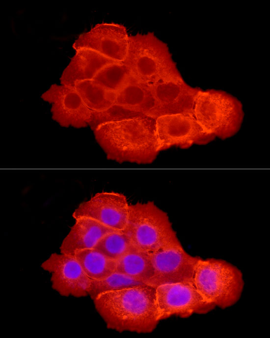 | Immunofluorescence analysis of A-431 cells using [KD Validated] EGFR Rabbit pAb (A11577) at dilution of 1:200 (40x lens). Secondary antibody: Cy3-conjugated Goat anti-Rabbit IgG (H+L) (AS007) at 1:500 dilution. Blue: DAPI for nuclear staining. |
You may also be interested in:

