| Reactivity: | Human |
| Applications: | WB, IF/ICC, ELISA |
| Host Species: | Rabbit |
| Isotype: | IgG |
| Clonality: | Monoclonal antibody |
| Gene Name: | glutathione peroxidase 4 |
| Gene Symbol: | GPX4 |
| Synonyms: | MCSP; SMDS; GPx-4; PHGPx; snGPx; GSHPx-4; snPHGPx; [KD Validated] GPX4 |
| Gene ID: | 2879 |
| UniProt ID: | P36969 |
| Clone ID: | 7O4Q9 |
| Immunogen: | A synthetic peptide corresponding to a sequence within amino acids 1-100 of human GPX4 (P36969). |
| Dilution: | WB 1:12500-1:50000; IHC 1:5000-1:20000; IF/IC 1:2000-1:8000 |
| Purification Method: | Affinity purification |
| Concentration: | 0.63 mg/mL |
| Buffer: | PBS with 0.09% sodium azid, 0.05% BSA, 50% glycerol, pH7.3. |
| Storage: | Store at -20°C. Avoid freeze / thaw cycles. |
| Documents: | Manual-GPX4 monoclonal antibody |
Background
The protein encoded by this gene belongs to the glutathione peroxidase family, members of which catalyze the reduction of hydrogen peroxide, organic hydroperoxides and lipid hydroperoxides, and thereby protect cells against oxidative damage. Several isozymes of this gene family exist in vertebrates, which vary in cellular location and substrate specificity. This isozyme has a high preference for lipid hydroperoxides and protects cells against membrane lipid peroxidation and cell death. It is also required for normal sperm development; thus, it has been identified as a 'moonlighting' protein because of its ability to serve dual functions as a peroxidase, as well as a structural protein in mature spermatozoa. Mutations in this gene are associated with Sedaghatian type of spondylometaphyseal dysplasia (SMDS). This isozyme is also a selenoprotein, containing the rare amino acid selenocysteine (Sec) at its active site. Sec is encoded by the UGA codon, which normally signals translation termination. The 3' UTRs of selenoprotein mRNAs contain a conserved stem-loop structure, designated the Sec insertion sequence (SECIS) element, that is necessary for the recognition of UGA as a Sec codon, rather than as a stop signal. Transcript variants resulting from alternative splicing or use of alternate promoters have been described to encode isoforms with different subcellular localization.
Images
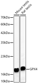 | Western blot analysis of various lysates using [KD Validated] GPX4 Rabbit mAb (A11243) at 1:1000 dilution incubated overnight at 4℃. Secondary antibody: HRP-conjugated Goat anti-Rabbit IgG (H+L) (AS014) at 1:10000 dilution. Lysates/proteins: 25 μg per lane. Blocking buffer: 3% nonfat dry milk in TBST. Detection: ECL Basic Kit (RM00020). Exposure time: 15s. |
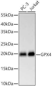 | Western blot analysis of various lysates using [KD Validated] GPX4 Rabbit mAb (A11243) at 1:1000 dilution incubated overnight at 4℃. Secondary antibody: HRP-conjugated Goat anti-Rabbit IgG (H+L) (AS014) at 1:10000 dilution. Lysates/proteins: 25 μg per lane. Blocking buffer: 3% nonfat dry milk in TBST. Detection: ECL Basic Kit (RM00020). Exposure time: 30s. |
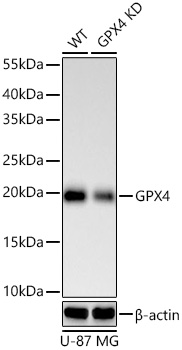 | Western blot analysis of lysates from wild type (WT) and GPX4 knockdown (KD) U-87 MG cells using [KD Validated] GPX4 Rabbit mAb (A11243) at 1:1000 dilution incubated overnight at 4℃. Secondary antibody: HRP-conjugated Goat anti-Rabbit IgG (H+L) (AS014) at 1:10000 dilution. Lysates/proteins: 25 μg per lane. Blocking buffer: 3% nonfat dry milk in TBST. Detection: ECL Basic Kit (RM00020). Exposure time: 60s. |
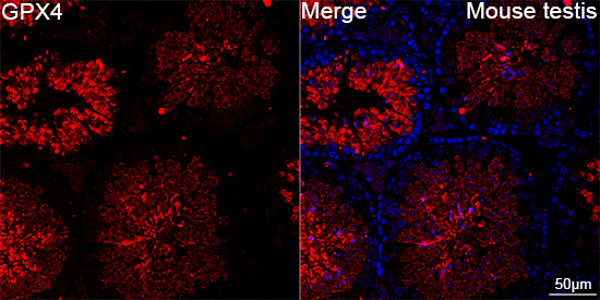 | Confocal imaging of paraffin-embedded Mouse testis tissue using [KD Validated] GPX4 Rabbit mAb (A11243, dilution 1:200) followed by a further incubation with Cy3 Goat Anti-Rabbit IgG (H+L) (AS007, dilution 1:500) (Red). DAPI was used for nuclear staining (Blue). High pressure antigen retrieval performed with 0.01M Citrate Buffer (pH 6.0) prior to IF staining. Objective: 40x. |
You may also be interested in:

