| Reactivity: | Human |
| Applications: | WB, IHC-P, IF/ICC, ELISA |
| Host Species: | Rabbit |
| Isotype: | IgG |
| Clonality: | Monoclonal antibody |
| Gene Name: | hydroxyacyl-CoA dehydrogenase trifunctional multienzyme complex subunit beta |
| Gene Symbol: | HADHB |
| Synonyms: | ECHB; MTPB; MTPD; MTPD2; MSTP029; TP-BETA; HADHB |
| Gene ID: | 3032 |
| UniProt ID: | P55084 |
| Clone ID: | 3N6Q10 |
| Immunogen: | Recombinant fusion protein containing a sequence corresponding to amino acids 331-474 of human HADHB(NP_000174.1). |
| Dilution: | WB 1:1000-1:5000; IF/IC 1:50-1:200 |
| Purification Method: | Affinity purification |
| Concentration: | 1.28 mg/mL |
| Buffer: | PBS with 0.05% proclin300, 0.05% BSA, 50% glycerol, pH7.3. |
| Storage: | Store at -20°C. Avoid freeze / thaw cycles. |
| Documents: | Manual-HADHB monoclonal antibody |
Background
This gene encodes the beta subunit of the mitochondrial trifunctional protein, which catalyzes the last three steps of mitochondrial beta-oxidation of long chain fatty acids. The mitochondrial membrane-bound heterocomplex is composed of four alpha and four beta subunits, with the beta subunit catalyzing the 3-ketoacyl-CoA thiolase activity. The encoded protein can also bind RNA and decreases the stability of some mRNAs. The genes of the alpha and beta subunits of the mitochondrial trifunctional protein are located adjacent to each other in the human genome in a head-to-head orientation. Mutations in this gene result in trifunctional protein deficiency. Alternatively spliced transcript variants encoding different isoforms have been described.
Images
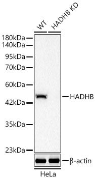 | Western blot analysis of lysates from wild type (WT) and HADHB knockdown (KD) HeLa cells using [KD Validated] HADHB Rabbit mAb (A25207) at 1:5000 dilution. Secondary antibody: HRP-conjugated Goat anti-Rabbit IgG (H+L) (AS014) at 1:10000 dilution. Lysates/proteins: 25 μg per lane. Blocking buffer: 3% nonfat dry milk in TBST. Detection: ECL Basic Kit (RM00020). Exposure time: 20s. |
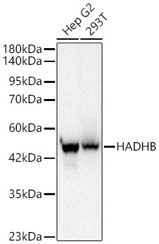 | Western blot analysis of various lysates using [KD Validated] HADHB Rabbit mAb (A25207) at 1:5000 dilution. Secondary antibody: HRP-conjugated Goat anti-Rabbit IgG (H+L) (AS014) at 1:10000 dilution. Lysates/proteins: 25 μg per lane. Blocking buffer: 3 % nonfat dry milk in TBST. Detection: ECL Basic Kit (RM00020). Exposure time: 20s. |
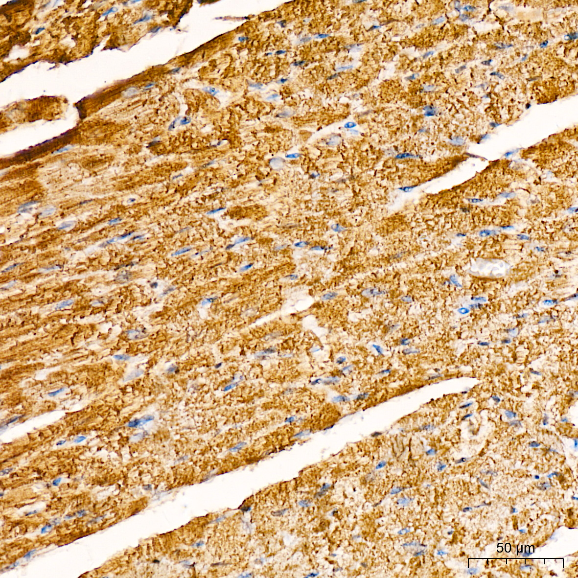 | Immunohistochemistry analysis of paraffin-embedded Mouse heart tissue using [KD Validated] HADHB Rabbit mAb (A25207) at a dilution of 1:200 (40x lens). High pressure antigen retrieval was performed with 0.01 M citrate buffer (pH 6.0) prior to IHC staining. |
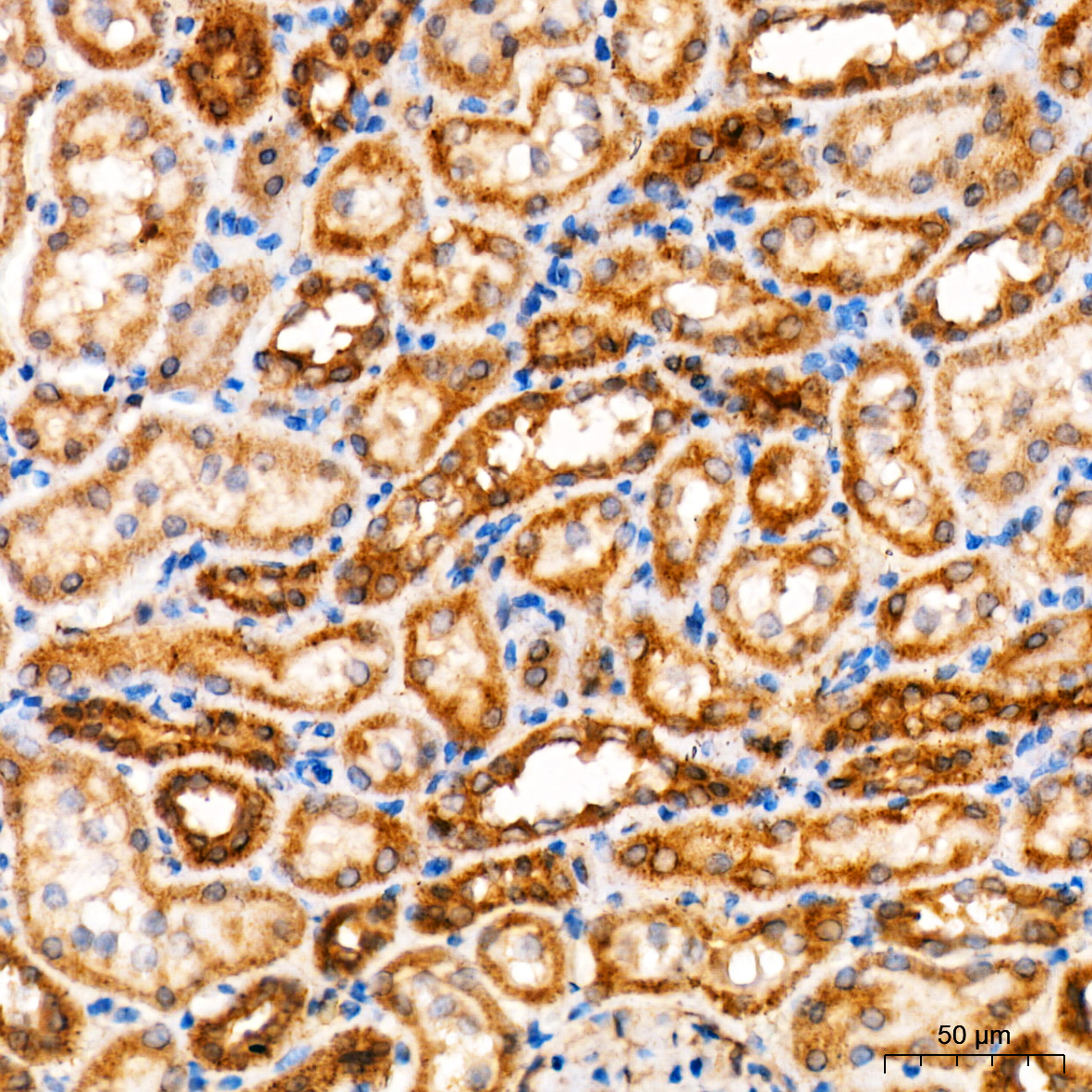 | Immunohistochemistry analysis of paraffin-embedded Rat kidney tissue using [KD Validated] HADHB Rabbit mAb (A25207) at a dilution of 1:200 (40x lens). High pressure antigen retrieval was performed with 0.01 M citrate buffer (pH 6.0) prior to IHC staining. |
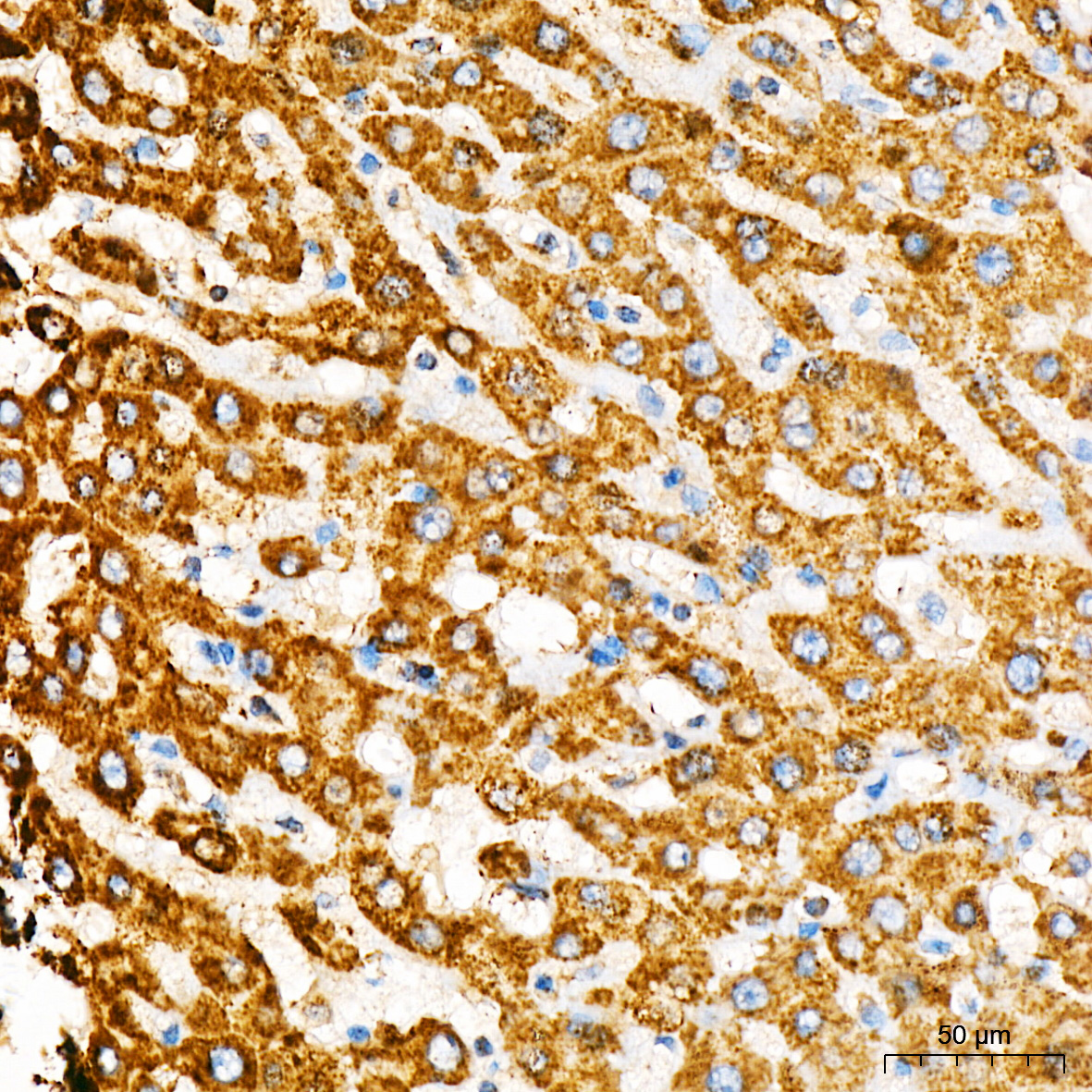 | Immunohistochemistry analysis of paraffin-embedded Human liver tissue using [KD Validated] HADHB Rabbit mAb (A25207) at a dilution of 1:200 (40x lens). High pressure antigen retrieval was performed with 0.01 M citrate buffer (pH 6.0) prior to IHC staining. |
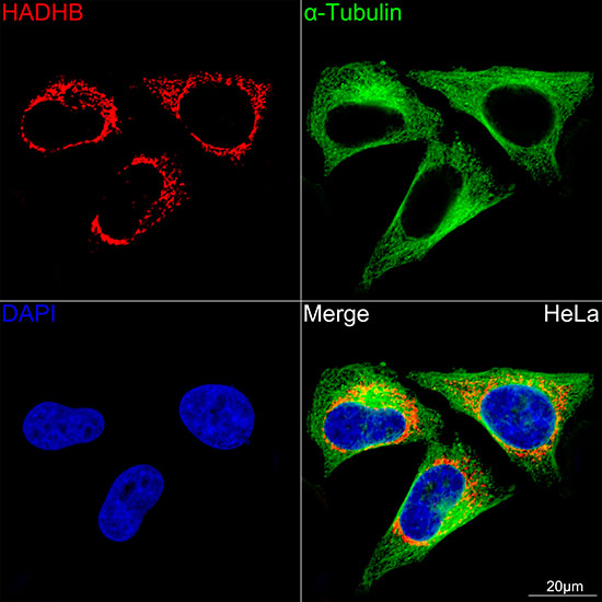 | Confocal imaging of HeLa cells using [KD Validated] HADHB Rabbit mAb (A25207, dilution 1:200) followed by a further incubation with Cy3 Goat Anti-Rabbit IgG (H+L) (AS007, dilution 1:500) (Red). The cells were counterstained with α-Tubulin Mouse mAb (AC012, dilution 1:400) followed by incubation with ABflo® 488-conjugated Goat Anti-Mouse IgG (H+L) Ab (AS076, dilution 1:500) (Green). DAPI was used for nuclear staining (Blue). Objective: 100x. |
You may also be interested in:

