Your shopping cart is empty!
")
KD-Validated LAMP1/CD107a Rabbit mAb (20 μl)
| Reactivity: | Human |
| Applications: | WB, IHC, IF/IC, FC (intra), ELISA |
| Host Species: | Rabbit |
| Isotype: | IgG |
| Clonality: | Monoclonal antibody |
| Gene Name: | lysosomal associated membrane protein 1 |
| Gene Symbol: | LAMP1 |
| Synonyms: | LAMPA; CD107a; LGP120 |
| Gene ID: | 3916 |
| UniProt ID: | P11279 |
| Clone ID: | 5O1C7 |
| Immunogen: | Recombinant fusion protein containing a sequence corresponding to amino acids 29-382 of human LAMP1/CD107a (NP_005552.3). |
| Dilution: | WB 1:10000-1:40000; IHC 1:500-1:5000; IF/IC 1:200-1:2000; FC (intra) 1:100-1:500 |
| Purification Method: | Affinity purification |
| Concentration: | 1 mg/ml |
| Buffer: | PBS with 0.05% proclin300, 0.05% BSA, 50% glycerol, pH7.3. |
| Storage: | Store at -20°C. Avoid freeze / thaw cycles. |
| Documents: | Manual-LAMP1 monoclonal antibody |
Background
The protein encoded by this gene is a member of a family of membrane glycoproteins. This glycoprotein provides selectins with carbohydrate ligands. It may also play a role in tumor cell metastasis.
Images
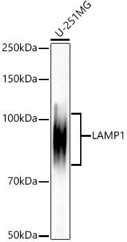 | Western blot analysis of lysates from U-251MG cells, using [KD Validated] LAMP1/CD107a Rabbit mAb (A21194) at 1:10000 dilution. Secondary antibody: HRP-conjugated Goat anti-Rabbit IgG (H+L) (AS014) at 1:10000 dilution. Lysates/proteins: 25μg per lane. Blocking buffer: 3% nonfat dry milk in TBST. Detection: ECL Basic Kit (RM00020). Exposure time: 20s. |
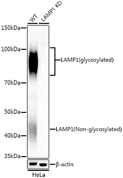 | Western blot analysis of lysates from wild type (WT) and LAMP1 knockdown (KD) HeLa cells, using [KD Validated] LAMP1/CD107a Rabbit mAb (A21194) at 1:10000 dilution. Secondary antibody: HRP-conjugated Goat anti-Rabbit IgG (H+L) (AS014) at 1:10000 dilution. Lysates/proteins: 25μg per lane. Blocking buffer: 3% nonfat dry milk in TBST. Detection: ECL Basic Kit (RM00020). Exposure time: 20s. |
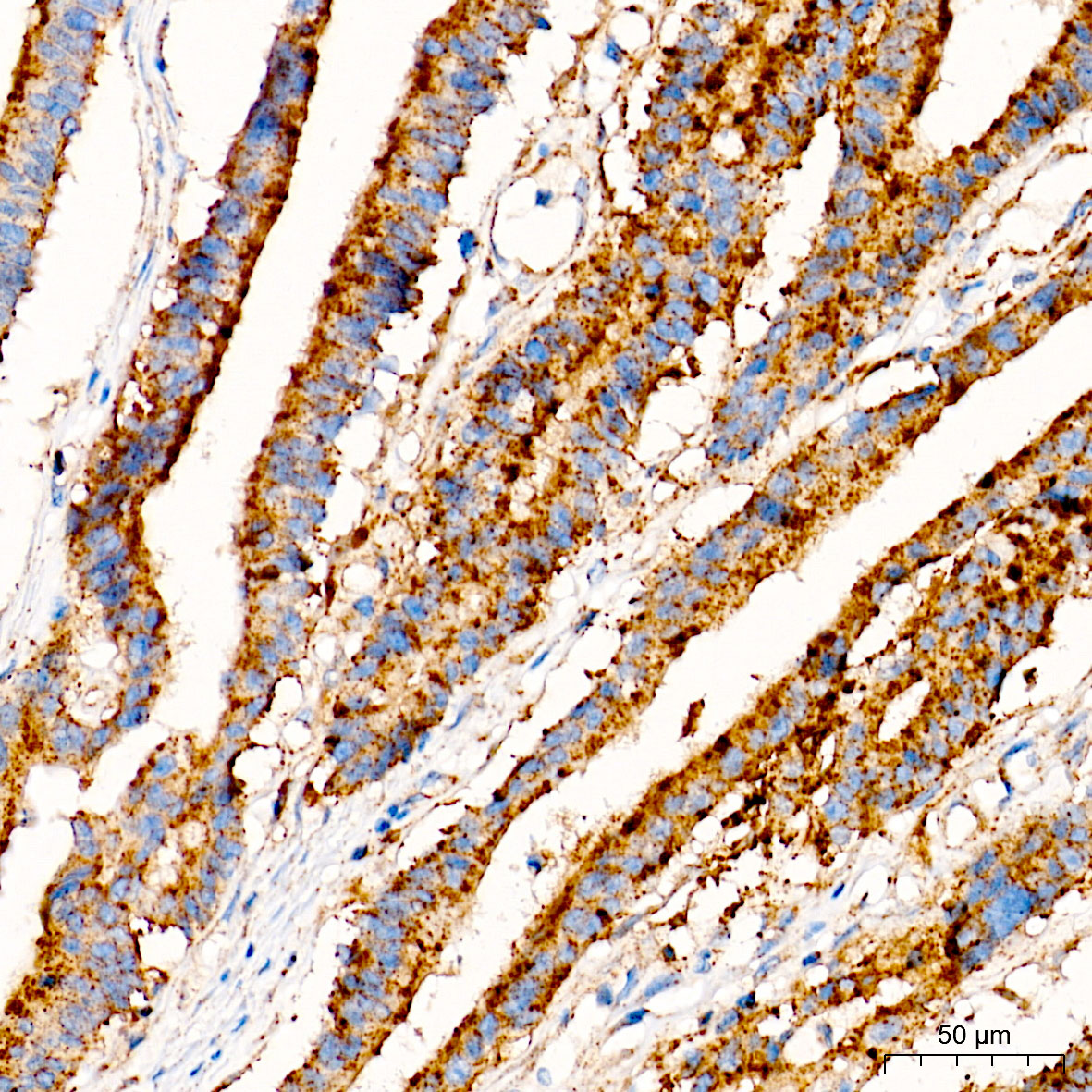 | Immunohistochemistry analysis of paraffin-embedded Human colon carcinoma tissue using [KD Validated] LAMP1/CD107a Rabbit mAb (A21194) at a dilution of 1:2000 (40x lens). High pressure antigen retrieval performed with 0.01M Tris-EDTA Buffer(pH 9.0) prior to IHC staining. |
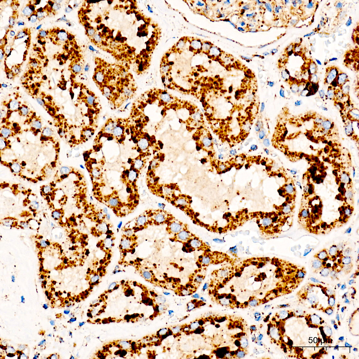 | Immunohistochemistry analysis of paraffin-embedded Human kidney tissue using [KD Validated] LAMP1/CD107a Rabbit mAb (A21194) at a dilution of 1:2000 (40x lens). High pressure antigen retrieval performed with 0.01M Tris-EDTA Buffer(pH 9.0) prior to IHC staining. |
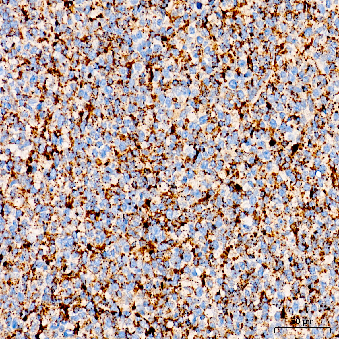 | Immunohistochemistry analysis of paraffin-embedded Human tonsil tissue using [KD Validated] LAMP1/CD107a Rabbit mAb (A21194) at a dilution of 1:2000 (40x lens). High pressure antigen retrieval performed with 0.01M Tris-EDTA Buffer(pH 9.0) prior to IHC staining. |
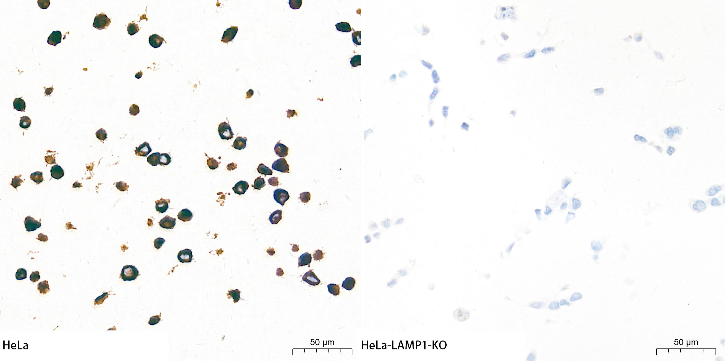 | Immunohistochemistry analysis of paraffin-embedded HeLa and HeLa-LAMP1-KO cells using [KD Validated] LAMP1/CD107a Rabbit mAb (A21194) at a dilution of 1:2200 (40x lens). High pressure antigen retrieval performed with 0.01M Tris-EDTA Buffer (pH 9.0) prior to IHC staining. |
 | Confocal imaging of HeLa and HeLa-LAMP1-KD cells using [KD Validated] LAMP1/CD107a Rabbit mAb (A21194,dilution 1:200) followed by a further incubation with Cy3 Goat Anti-Rabbit IgG (H+L) (AS007,dilution 1:500)(Red).DAPI was used for nuclear staining (Blue). Objective: 100x. |
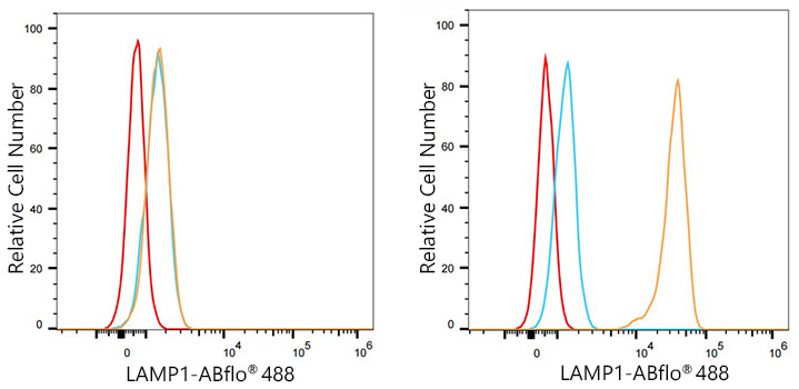 | Flow cytometry: 1X10^6 knockdown (KD) HeLa cells (negative control,left) and HeLa cells (right) were intracellularly-stained with [KD Validated] LAMP1/CD107a Rabbit mAb (A21194,2.5 μg/mL,orange line) or ABflo® 488 Rabbit IgG isotype control (A22069,5 μl/Test,blue line), followed by FITC conjugated goat anti-Rabbit pAb staining. Non-fluorescently stained cells were used as blank control (red line). |
You may also be interested in:

