KO-Validated APEX1/APE1 Rabbit pAb (20 μl)
| Reactivity: | Human |
| Applications: | WB, IF/IC, IP, ELISA |
| Host Species: | Rabbit |
| Isotype: | IgG |
| Clonality: | Polyclonal antibody |
| Gene Name: | apurinic/apyrimidinic endodeoxyribonuclease 1 |
| Gene Symbol: | APEX1 |
| Synonyms: | APE; APX; APE1; APEN; APEX; HAP1; REF1; E1 |
| Gene ID: | 328 |
| UniProt ID: | P27695 |
| Immunogen: | Recombinant fusion protein containing a sequence corresponding to amino acids 1-318 of human APEX1/APE1 (NP_542380.1). |
| Dilution: | WB 1:500-1:2000; IF/IC 1:50-1:100 |
| Purification Method: | Affinity purification |
| Concentration: | 2.29 mg/ml |
| Buffer: | PBS with 0.02% sodium azide, 50% glycerol ,pH7.3. |
| Storage: | Store at -20°C. Avoid freeze / thaw cycles. |
| Documents: | Manual-APEX1 polyclonal antibody |
Background
The APEX gene encodes the major AP endonuclease in human cells. It encodes the APEX endonuclease, a DNA repair enzyme with apurinic/apyrimidinic (AP) activity. Such AP activity sites occur frequently in DNA molecules by spontaneous hydrolysis, by DNA damaging agents or by DNA glycosylases that remove specific abnormal bases. The AP sites are the most frequent pre-mutagenic lesions that can prevent normal DNA replication. Splice variants have been found for the gene APEX1; all encode the same protein. Disruptions in the biological functions related to APEX are associated with many various malignancies and neurodegenerative diseases.
Images
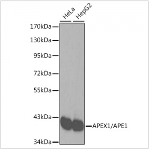
 | Western blot analysis of various lysates using [KO Validated] APEX1/APE1 Rabbit pAb (A2587). Secondary antibody: HRP-conjugated Goat anti-Rabbit IgG (H+L) (AS014) at 1:10000 dilution. Lysates/proteins: 25μg per lane. Blocking buffer: 3% nonfat dry milk in TBST. |
 | Western blot analysis of lysates from wild type (WT) and APEX1/APE1 knockout (KO) 293T cells, using [KO Validated] APEX1/APE1 Rabbit pAb (A2587) at 1:3000 dilution. Secondary antibody: HRP-conjugated Goat anti-Rabbit IgG (H+L) (AS014) at 1:10000 dilution. Lysates/proteins: 25μg per lane. Blocking buffer: 3% nonfat dry milk in TBST. Detection: ECL Basic Kit (RM00020). Exposure time: 1s. |
 | Immunofluorescence analysis of HeLa cells using [KO Validated] APEX1/APE1 Rabbit pAb (A2587). Secondary antibody: Cy3-conjugated Goat anti-Rabbit IgG (H+L) (AS007) at 1:500 dilution. Blue: DAPI for nuclear staining. |
 | Immunofluorescence analysis of A549 cells using [KO Validated] APEX1/APE1 Rabbit pAb (A2587).Secondary antibody: Cy3-conjugated Goat anti-Rabbit IgG (H+L) (AS007) at 1:500 dilution. |
 | Immunoprecipitation analysis of 200 μg extracts of HeLa cells using 1 μg APEX1/APE1 antibody (A2587). Western blot was performed from the immunoprecipitate using APEX1/APE1 antibody (A2587) at a dilution of 1:1000. |
You may also be interested in:


