KO-Validated Caspase-3 Rabbit mAb (20 μl)
| Reactivity: | Human |
| Applications: | WB, ELISA |
| Host Species: | Rabbit |
| Isotype: | IgG |
| Clonality: | Monoclonal antibody |
| Gene Name: | caspase 3 |
| Gene Symbol: | CASP3 |
| Synonyms: | CPP32; SCA-1; CPP32B; -3 |
| Gene ID: | 836 |
| UniProt ID: | P42574 |
| Clone ID: | 7T1W5 |
| Immunogen: | Recombinant fusion protein containing a sequence corresponding to amino acids 29-175 of human Caspase-3 (P42574). |
| Dilution: | WB 1:1000-1:2000; IHC 1:200-1:800 |
| Purification Method: | Affinity purification |
| Buffer: | PBS with 0.09% sodium azid, 0.05% BSA, 50% glycerol, pH7.3. |
| Storage: | Store at -20°C. Avoid freeze / thaw cycles. |
| Documents: | Manual-CASP3 monoclonal antibody |
Background
The protein encoded by this gene is a cysteine-aspartic acid protease that plays a central role in the execution-phase of cell apoptosis. The encoded protein cleaves and inactivates poly(ADP-ribose) polymerase while it cleaves and activates sterol regulatory element binding proteins as well as caspases 6, 7, and 9. This protein itself is processed by caspases 8, 9, and 10. It is the predominant caspase involved in the cleavage of amyloid-beta 4A precursor protein, which is associated with neuronal death in Alzheimer's disease.
Images
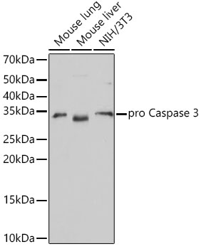
 | Western blot analysis of various lysates, using [KO Validated] active + pro Caspase-3 Rabbit mAb (A19654) at 1:1000 dilution. Secondary antibody: HRP-conjugated Goat anti-Rabbit IgG (H+L) (AS014) at 1:10000 dilution. Lysates/proteins: 25μg per lane. Blocking buffer: 3% nonfat dry milk in TBST. Detection: ECL Basic Kit (RM00020). Exposure time: 3min. |
 | Western blot analysis of lysates from Jurkat cells, using [KO Validated] active + pro Caspase-3 Rabbit mAb (A19654) at 1:1000 dilution. Jurkat cells were treated by Etoposide (25 uM) at 37℃ for 5 hours. Secondary antibody: HRP-conjugated Goat anti-Rabbit IgG (H+L) (AS014) at 1:10000 dilution. Lysates/proteins: 25μg per lane. Blocking buffer: 3% nonfat dry milk in TBST. Detection: ECL Basic Kit (RM00020). Exposure time: 90s. |
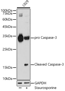
 | Western blot analysis of lysates from L929 cells, using [KO Validated] active + pro Caspase-3 Rabbit mAb (A19654) at 1:1000 dilution. L929 cells were treated by staurosporine(1 uM) for 3 hour. Secondary antibody: HRP-conjugated Goat anti-Rabbit IgG (H+L) (AS014) at 1:10000 dilution. Lysates/proteins: 25μg per lane. Blocking buffer: 3% nonfat dry milk in TBST. Detection: ECL Basic Kit (RM00020). Exposure time: 180s. |
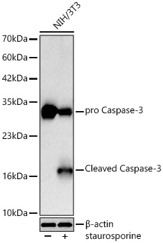
 | Western blot analysis of lysates from NIH/3T3 cells using Caspase-3 Rabbit mAb (A19654) at 1:1000 dilution incubated overnight at 4℃. NIH/3T3 cells were treated by staurosporine(1 μM) for 3 hour. Secondary antibody: HRP-conjugated Goat anti-Rabbit IgG (H+L) (AS014) at 1:10000 dilution. Lysates/proteins: 30 μg per lane. Blocking buffer: 3 % nonfat dry milk in TBST. Detection: ECL Basic Kit (RM00020). Exposure time: 90s. |
 | Western blot analysis of lysates from wild type (WT) and Caspase-3 knockout (KO) 293T cells using [KO Validated] Caspase-3 Rabbit mAb (A19654) at 1:1000 dilution incubated overnight at 4℃. Secondary antibody: HRP-conjugated Goat anti-Rabbit IgG (H+L) (AS014) at 1:10000 dilution. Lysates/proteins: 25 μg per lane. Blocking buffer: 3% nonfat dry milk in TBST. Detection: ECL Basic Kit (RM00020). Exposure time: 10s. |
You may also be interested in:


