KO-Validated ErbB3/HER3 Rabbit pAb (20 μl)
| Reactivity: | Human |
| Applications: | WB, IF/ICC, ELISA |
| Host Species: | Rabbit |
| Isotype: | IgG |
| Clonality: | Polyclonal antibody |
| Gene Name: | erb-b2 receptor tyrosine kinase 3 |
| Gene Symbol: | ERBB3 |
| Synonyms: | HER3; FERLK; LCCS2; VSCN1; ErbB-3; c-erbB3; erbB3-S; MDA-BF-1; c-erbB-3; p180-ErbB3; p45-sErbB3; p85-sErbB3; R3 |
| Gene ID: | 2065 |
| UniProt ID: | P21860 |
| Immunogen: | Recombinant fusion protein containing a sequence corresponding to amino acids 1275-1342 of human ErbB3/HER3 (NP_001973.2). |
| Dilution: | WB 1:1000-1:6000; IF/IC 1:200-1:800 |
| Purification Method: | Affinity purification |
| Concentration: | 2.31 mg/ml |
| Buffer: | PBS with 0.02% sodium azide, 50% glycerol ,pH7.3. |
| Storage: | Store at -20°C. Avoid freeze / thaw cycles. |
| Documents: | Manual-ERBB3 polyclonal antibody |
Background
This gene encodes a member of the epidermal growth factor receptor (EGFR) family of receptor tyrosine kinases. This membrane-bound protein has a neuregulin binding domain but not an active kinase domain. It therefore can bind this ligand but not convey the signal into the cell through protein phosphorylation. However, it does form heterodimers with other EGF receptor family members which do have kinase activity. Heterodimerization leads to the activation of pathways which lead to cell proliferation or differentiation. Amplification of this gene and/or overexpression of its protein have been reported in numerous cancers, including prostate, bladder, and breast tumors. Alternate transcriptional splice variants encoding different isoforms have been characterized. One isoform lacks the intermembrane region and is secreted outside the cell. This form acts to modulate the activity of the membrane-bound form. Additional splice variants have also been reported, but they have not been thoroughly characterized.
Images
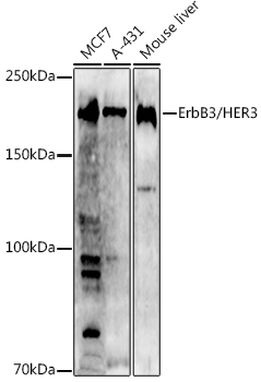
 | Western blot analysis of various lysates using [KO Validated] ErbB3/HER3 Rabbit pAb (A0950) at 1:1000 dilution. Secondary antibody: HRP-conjugated Goat anti-Rabbit IgG (H+L) (AS014) at 1:10000 dilution. Lysates/proteins: 25μg per lane. Blocking buffer: 3% nonfat dry milk in TBST. Detection: ECL Basic Kit (RM00020). Exposure time: 180s. |
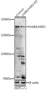
 | Western blot analysis of lysates from wild type (WT) and ErbB3/HER3 knockout (KO) 293T cells, using [KO Validated] ErbB3/HER3 Rabbit pAb (A0950) at 1:1000 dilution. Secondary antibody: HRP-conjugated Goat anti-Rabbit IgG (H+L) (AS014) at 1:10000 dilution. Lysates/proteins: 25μg per lane. Blocking buffer: 3% nonfat dry milk in TBST. Detection: ECL Basic Kit (RM00020). Exposure time: 180s. |
You may also be interested in:


