Your shopping cart is empty!
")
KO-Validated Hsp90α Rabbit mAb (20 μl)
| Reactivity: | Human, Mouse, Rat |
| Applications: | WB, IF/IC, ELISA |
| Host Species: | Rabbit |
| Isotype: | IgG |
| Clonality: | Monoclonal antibody |
| Gene Name: | heat shock protein 90 alpha family class A member 1 |
| Gene Symbol: | HSP90AA1 |
| Synonyms: | EL52; HSPN; LAP2; HSP86; HSPC1; HSPCA; Hsp89; Hsp90; LAP-2; HSP89A; HSP90A; HSP90N; Hsp103; HSPCAL1; HSPCAL4; HEL-S-65p |
| Gene ID: | 3320 |
| UniProt ID: | P07900 |
| Immunogen: | Recombinant fusion protein containing a sequence corresponding to amino acids 433-732 of human Hsp90α (NP_005339.3). |
| Dilution: | WB 1:5000-1:20000; IF/IC 1:200-1:800 |
| Purification Method: | Affinity purification |
| Concentration: | 2.02 mg/ml |
| Buffer: | PBS with 0.09% Sodium azide, 0.05% BSA, 50% glycerol, pH7.3. |
| Storage: | Store at -20°C. Avoid freeze / thaw cycles. |
| Documents: | Manual-HSP90AA1 monoclonal antibody |
Background
The protein encoded by this gene is an inducible molecular chaperone that functions as a homodimer. The encoded protein aids in the proper folding of specific target proteins by use of an ATPase activity that is modulated by co-chaperones. Two transcript variants encoding different isoforms have been found for this gene.
Images
 | Western blot analysis of various lysates using [KO Validated] Hsp90α Rabbit mAb (A26677) at 1:5000 dilution incubated overnight at 4℃. Secondary antibody: HRP-conjugated Goat anti-Rabbit IgG (H+L) (AS014) at 1:10000 dilution. Lysates/proteins: 25 μg per lane. Blocking buffer: 3% nonfat dry milk in TBST. Detection: ECL Basic Kit (RM00020). Exposure time: 30s. |
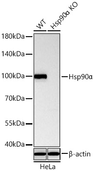 | Western blot analysis of lysates from wild type (WT) and Hsp90α knockout (KO) HeLa cells using [KO Validated] Hsp90α Rabbit mAb (A26677) at 1:5000 dilution incubated overnight at 4℃. Secondary antibody: HRP-conjugated Goat anti-Rabbit IgG (H+L) (AS014) at 1:10000 dilution. Lysates/proteins: 25 μg per lane. Blocking buffer: 3% nonfat dry milk in TBST. Detection: ECL Basic Kit (RM00020). Exposure time: 30s. |
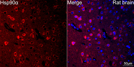 | Confocal imaging of paraffin-embedded Rat brain tissue using [KO Validated] Hsp90α Rabbit mAb (A26677, dilution 1:200) followed by a further incubation with Cy3 Goat Anti-Rabbit IgG (H+L) (AS007, dilution 1:500) (Red). DAPI was used for nuclear staining (Blue). Microwave antigen retrieval performed with 0.01M Citrate Buffer(pH 6.0) prior to IF staining. Objective: 40x. |
 | Confocal imaging of paraffin-embedded Mouse brain tissue using [KO Validated] Hsp90α Rabbit mAb (A26677, dilution 1:200) followed by a further incubation with Cy3 Goat Anti-Rabbit IgG (H+L) (AS007, dilution 1:500) (Red). DAPI was used for nuclear staining (Blue). Microwave antigen retrieval performed with 0.01M Citrate Buffer(pH 6.0) prior to IF staining. Objective: 40x. |
You may also be interested in:

