Your shopping cart is empty!
")
| Reactivity: | Human, Mouse, Rat |
| Applications: | WB, IHC, IF/IC, IP, ELISA |
| Host Species: | Rabbit |
| Isotype: | IgG |
| Clonality: | Monoclonal antibody |
| Gene Name: | interferon regulatory factor 3 |
| Gene Symbol: | IRF3 |
| Synonyms: | IIAE7 |
| Gene ID: | 3661 |
| UniProt ID: | Q14653 |
| Immunogen: | Recombinant fusion protein containing a sequence corresponding to amino acids 1-260 of human IRF3 (NP_001184051.1). |
| Dilution: | WB 1:11000-1:44000; IHC 1:600-1:3000; IF/IC 1:100-1:400 |
| Purification Method: | Affinity purification |
| Concentration: | 1.13 mg/ml |
| Buffer: | PBS with 0.09% Sodium azide, 0.05% BSA, 50% glycerol, pH7.3. |
| Storage: | Store at -20°C. Avoid freeze / thaw cycles. |
| Documents: | Manual-IRF3 monoclonal antibody |
Background
This gene encodes a member of the interferon regulatory transcription factor (IRF) family. The encoded protein is found in an inactive cytoplasmic form that upon serine/threonine phosphorylation forms a complex with CREBBP. This complex translocates to the nucleus and activates the transcription of interferons alpha and beta, as well as other interferon-induced genes. The protein plays an important role in the innate immune response against DNA and RNA viruses. Mutations in this gene are associated with Encephalopathy, acute, infection-induced, herpes-specific, 7.
Images
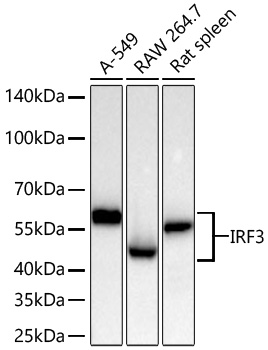 | Western blot analysis of various lysates using [KO Validated] IRF3 Rabbit mAb (A27272) at 1:11000 dilution incubated overnight at 4℃. Secondary antibody: HRP-conjugated Goat anti-Rabbit IgG (H+L) (AS014) at 1:10000 dilution. Lysates/proteins: 25 μg per lane. Blocking buffer: 3% nonfat dry milk in TBST. Detection: ECL Basic Kit (RM00020). Exposure time: 30s. |
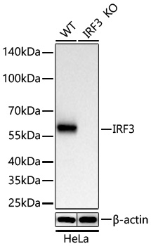 | Western blot analysis of lysates from wild type (WT) and IRF3 knockout (KO) HeLa cells using [KO Validated] IRF3 Rabbit mAb (A27272) at 1:11000 dilution incubated overnight at 4℃. Secondary antibody: HRP-conjugated Goat anti-Rabbit IgG (H+L) (AS014) at 1:10000 dilution. Lysates/proteins: 25 μg per lane. Blocking buffer: 3% nonfat dry milk in TBST. Detection: ECL Basic Kit (RM00020). Exposure time: 30s. |
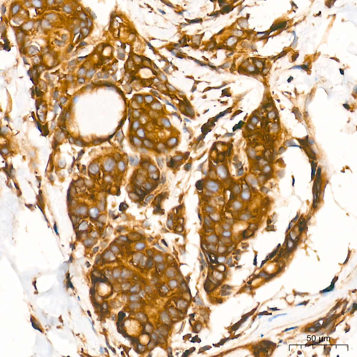
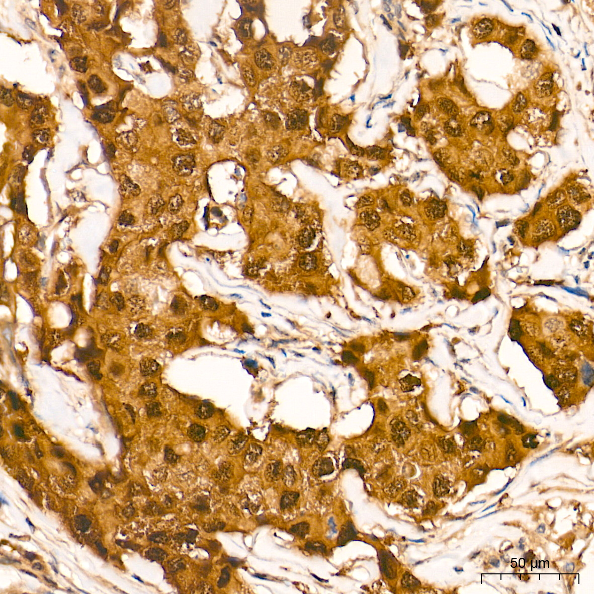 | Immunohistochemistry analysis of paraffin-embedded Human breast cancer tissue using [KO Validated] IRF3 Rabbit mAb (A27272) at a dilution of 1:2000 (40x lens). High pressure antigen retrieval performed with 0.01M Tris-EDTA Buffer (pH 9.0) prior to IHC staining. |
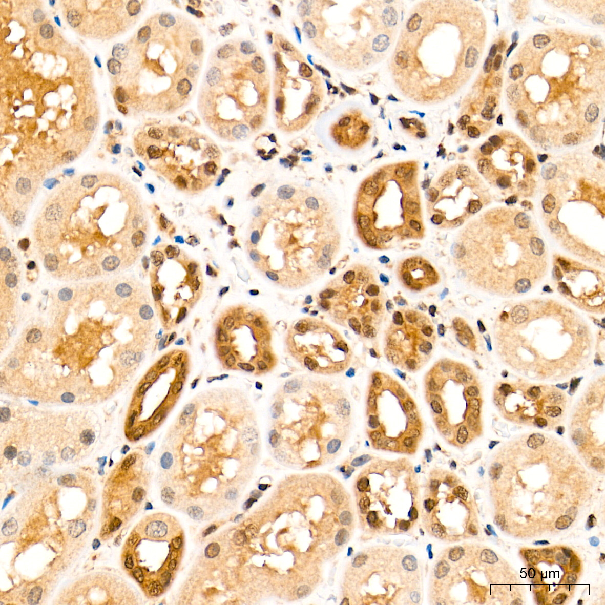 | Immunohistochemistry analysis of paraffin-embedded Human kidney tissue using [KO Validated] IRF3 Rabbit mAb (A27272) at a dilution of 1:2000 (40x lens). High pressure antigen retrieval performed with 0.01M Tris-EDTA Buffer (pH 9.0) prior to IHC staining. |
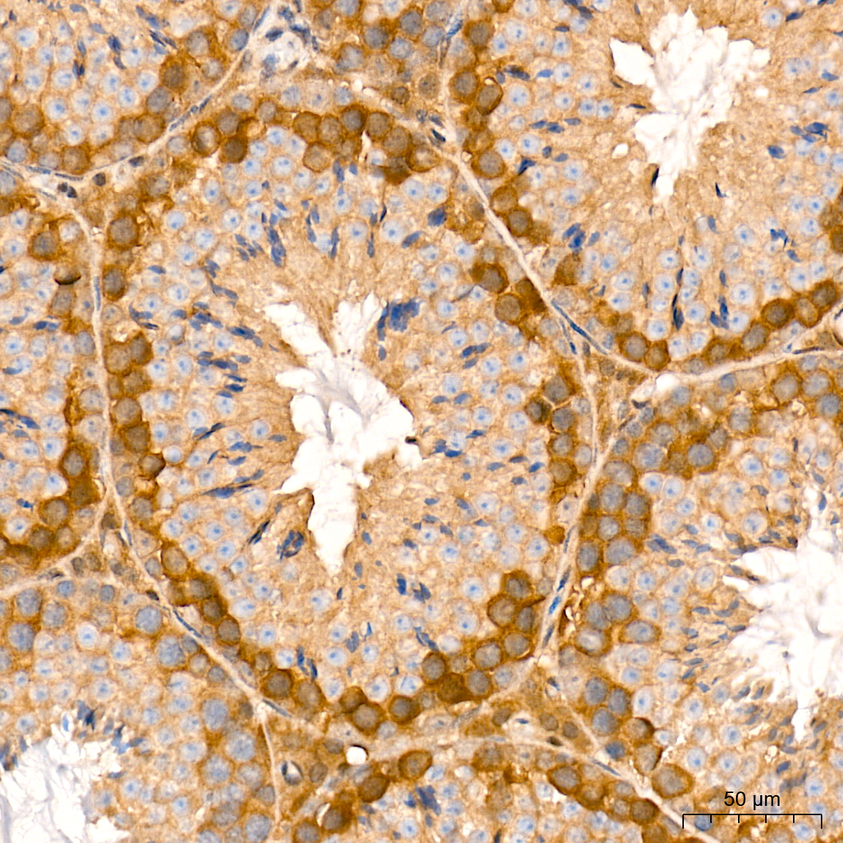
 | Immunohistochemistry analysis of paraffin-embedded Human thyroid cancer tissue using [KO Validated] IRF3 Rabbit mAb (A27272) at a dilution of 1:2000 (40x lens). High pressure antigen retrieval performed with 0.01M Tris-EDTA Buffer (pH 9.0) prior to IHC staining. |
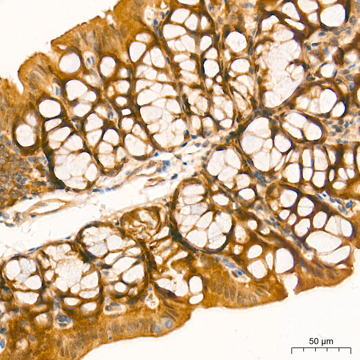 | Immunohistochemistry analysis of paraffin-embedded Mouse intestin tissue using [KO Validated] IRF3 Rabbit mAb (A27272) at a dilution of 1:2000 (40x lens). High pressure antigen retrieval performed with 0.01M Tris-EDTA Buffer (pH 9.0) prior to IHC staining. |
 | Immunohistochemistry analysis of paraffin-embedded Mouse testis tissue using [KO Validated] IRF3 Rabbit mAb (A27272) at a dilution of 1:2000 (40x lens). High pressure antigen retrieval performed with 0.01M Tris-EDTA Buffer (pH 9.0) prior to IHC staining. |
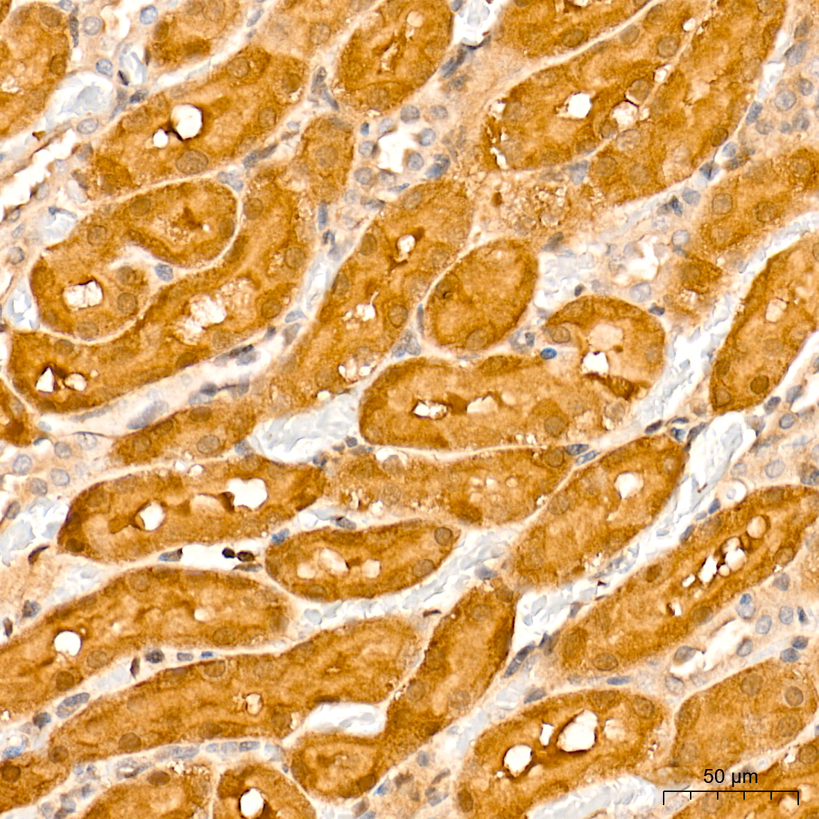 | Immunohistochemistry analysis of paraffin-embedded Rat kidney tissue using [KO Validated] IRF3 Rabbit mAb (A27272) at a dilution of 1:2000 (40x lens). High pressure antigen retrieval performed with 0.01M Tris-EDTA Buffer (pH 9.0) prior to IHC staining. |
You may also be interested in:

