Your shopping cart is empty!
")
| Reactivity: | Human, Mouse, Rat |
| Applications: | WB, IF/IC, ELISA |
| Host Species: | Rabbit |
| Isotype: | IgG |
| Clonality: | Monoclonal antibody |
| Gene Name: | actate dehydrogenase A |
| Gene Symbol: | LDHA |
| Synonyms: | LDHM; GSD11; PIG19; HEL-S-133P; HA |
| Gene ID: | 3939 |
| UniProt ID: | P00338 |
| Clone ID: | 5U6N0 |
| Immunogen: | A synthetic peptide corresponding to a sequence within amino acids 200-300 of human LDHA (P00338). |
| Dilution: | WB 1:1000-1:2000; IF/IC 1:100-1:800 |
| Purification Method: | Affinity purification |
| Concentration: | 0.8 mg/mL |
| Buffer: | PBS with 0.02% sodium azide, 0.05% BSA, 50% glycerol, pH7.3. |
| Storage: | Store at -20°C. Avoid freeze / thaw cycles. |
| Documents: | Manual-LDHA monoclonal antibody |
Background
The protein encoded by this gene catalyzes the conversion of L-lactate and NAD to pyruvate and NADH in the final step of anaerobic glycolysis. The protein is found predominantly in muscle tissue and belongs to the lactate dehydrogenase family. Mutations in this gene have been linked to exertional myoglobinuria. Multiple transcript variants encoding different isoforms have been found for this gene. The human genome contains several non-transcribed pseudogenes of this gene.
Images
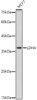 | Western blot analysis of lysates from MCF7 cells, using [KO Validated] LDHA Rabbit mAb (A0861) at 1:1000 dilution. Secondary antibody: HRP-conjugated Goat anti-Rabbit IgG (H+L) (AS014) at 1:10000 dilution. Lysates/proteins: 25μg per lane. Blocking buffer: 3% nonfat dry milk in TBST. Detection: ECL Basic Kit (RM00020). Exposure time: 180s. |
 | Western blot analysis of lysates from wild type(WT) and LDHA knockout (KO) 293T cells, using [KO Validated] LDHA Rabbit mAb (A0861) at 1:1000 dilution. Secondary antibody: HRP-conjugated Goat anti-Rabbit IgG (H+L) (AS014) at 1:10000 dilution. Lysates/proteins: 25μg per lane. Blocking buffer: 3% nonfat dry milk in TBST. Detection: ECL Basic Kit (RM00020). Exposure time: 180s. |
 | Immunofluorescence analysis of NIH/3T3 cells using [KO Validated] LDHA Rabbit mAb (A0861) at dilution of 1:100 (40x lens). Secondary antibody: Cy3-conjugated Goat anti-Rabbit IgG (H+L) (AS007) at 1:500 dilution. Blue: DAPI for nuclear staining. |
 | Immunofluorescence analysis of PC-12 cells using [KO Validated] LDHA Rabbit mAb (A0861) at dilution of 1:100 (40x lens). Secondary antibody: Cy3-conjugated Goat anti-Rabbit IgG (H+L) (AS007) at 1:500 dilution. Blue: DAPI for nuclear staining. |
 | Immunofluorescence analysis of U2OS cells using [KO Validated] LDHA Rabbit mAb (A0861) at dilution of 1:100 (40x lens). Secondary antibody: Cy3-conjugated Goat anti-Rabbit IgG (H+L) (AS007) at 1:500 dilution. Blue: DAPI for nuclear staining. |
You may also be interested in:

