Your shopping cart is empty!
")
KO-Validated Lamin A/C Rabbit mAb (20 μl)
| Reactivity: | Human, Mouse, Rat |
| Applications: | WB, IHC, IF/IC, IP, ELISA |
| Host Species: | Rabbit |
| Isotype: | IgG |
| Clonality: | Monoclonal antibody |
| Gene Name: | lamin A/C |
| Gene Symbol: | LMNA |
| Synonyms: | FPL; IDC; LFP; CDDC; EMD2; FPLD; HGPS; LDP1; LMN1; LMNC; MADA; PRO1; CDCD1; CMD1A; FPLD2; LMNL1; CMT2B1; LGMD1B; /C |
| Gene ID: | 4000 |
| UniProt ID: | P02545 |
| Clone ID: | 4L8Q0 |
| Immunogen: | Recombinant fusion protein containing a sequence corresponding to amino acids 403-572 of human Lamin A/C (NP_733821.1). |
| Dilution: | WB 1:50000-1:300000; IHC 1:1000-1:4000; IF/IC 1:100-1:800 |
| Purification Method: | Affinity purification |
| Concentration: | 1.2mg/mL |
| Buffer: | PBS with 0.05% proclin300, 0.05% BSA, 50% glycerol, pH7.3. |
| Storage: | Store at -20°C. Avoid freeze / thaw cycles. |
| Documents: | Manual-LMNA monoclonal antibody |
Background
The protein encoded by this gene is part of the nuclear lamina, a two-dimensional matrix of proteins located next to the inner nuclear membrane. The lamin family of proteins make up the matrix and are highly conserved in evolution. During mitosis, the lamina matrix is reversibly disassembled as the lamin proteins are phosphorylated. Lamin proteins are thought to be involved in nuclear stability, chromatin structure and gene expression. Vertebrate lamins consist of two types, A and B. Alternative splicing results in multiple transcript variants. Mutations in this gene lead to several diseases: Emery-Dreifuss muscular dystrophy, familial partial lipodystrophy, limb girdle muscular dystrophy, dilated cardiomyopathy, Charcot-Marie-Tooth disease, and Hutchinson-Gilford progeria syndrome.
Images
 | The STORM super-resolution (SR) imaging of U-2 OS cells using [KO Validated] Lamin A/C Rabbit mAb (A19524, ABclonal) at dilution of 1:200 with 3% paraformaldehyde (PFA) +0.1% glutaraldehyde (GA) fixation. The immunostaining was performed by Full Automatic Immunofluorescence Workflow System (Workflow Ultra300, Nano-Micro imaging, China). Image was performed with Single-Molecule Localization Super-Resolution Microscopy (STORM Ultra300, Nano-Micro imaging, China). We acknowledge Nano-Micro imaging Biotechnology Co., Ltd. (宁波纳微成像生物科技有限公司) in SR image processing and kindly providing this image. |
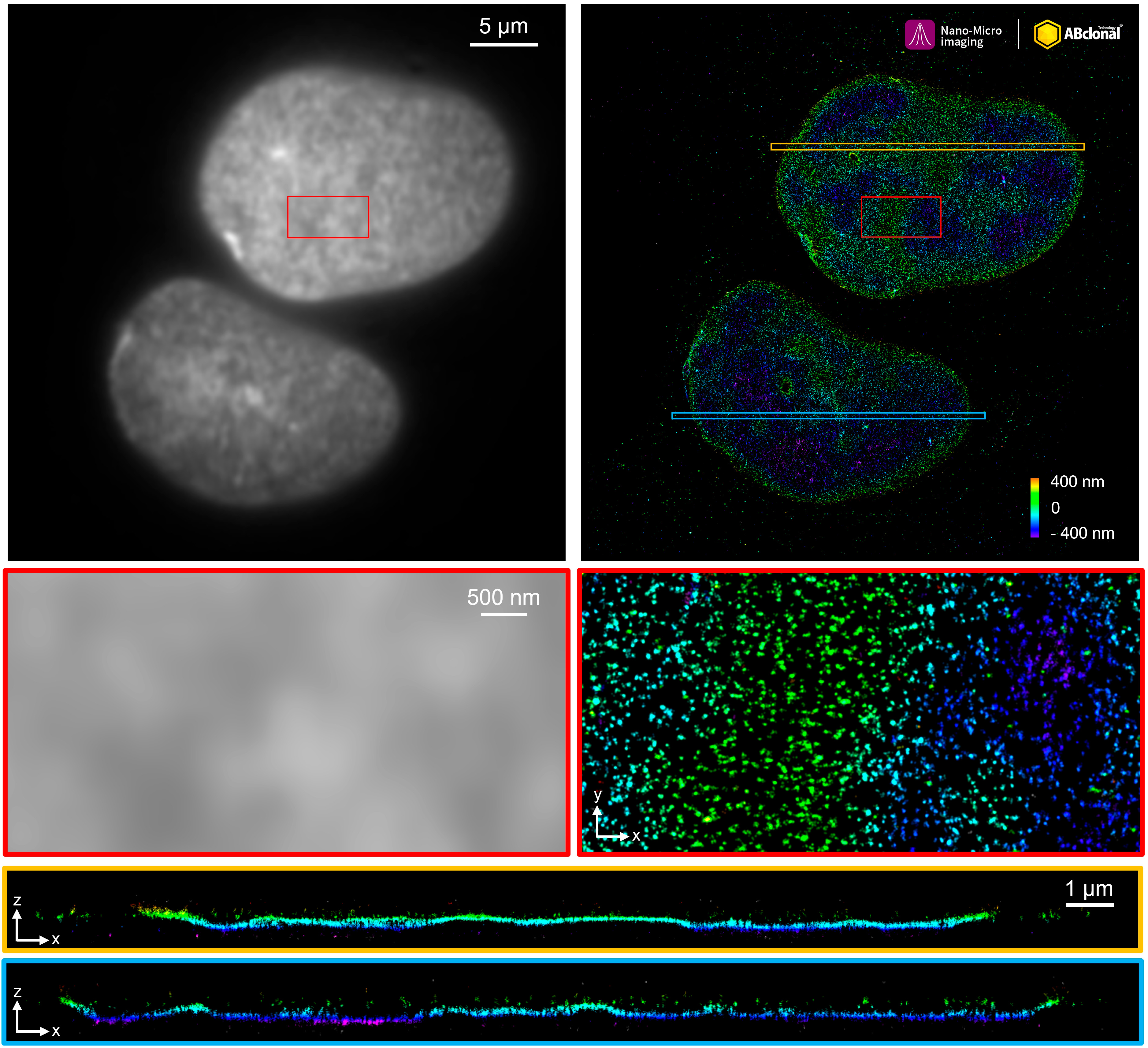 | The STORM super-resolution (SR) imaging of U-2 OS cells using [KO Validated] Lamin A/C Rabbit mAb (A19524, ABclonal) at dilution of 1:200 with 3% paraformaldehyde (PFA) +0.1% glutaraldehyde (GA) fixation. The immunostaining was performed by Full Automatic Immunofluorescence Workflow System (Workflow Ultra300, Nano-Micro imaging, China). Image was performed with Single-Molecule Localization Super-Resolution Microscopy (STORM Ultra300, Nano-Micro imaging, China). We acknowledge Nano-Micro imaging Biotechnology Co., Ltd. in SR image processing and kindly providing this image. |
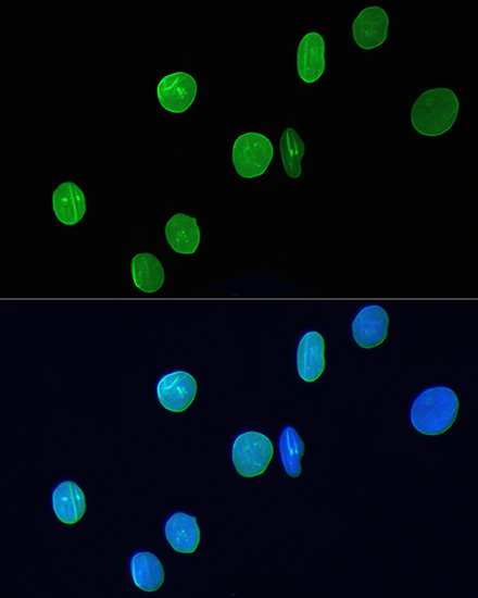
 | Immunofluorescence analysis of H9C2 cells using [KO Validated] Lamin A/C Rabbit mAb (A19524) at dilution of 1:200. Blue: DAPI for nuclear staining. |
 | Immunofluorescence analysis of L929 cells using [KO Validated] Lamin A/C Rabbit mAb (A19524) at dilution of 1:200. Blue: DAPI for nuclear staining. |
 | Confocal immunofluorescence analysis of HeLa cells using [KO Validated] Lamin A/C Rabbit mAb (A19524) at dilution of 1:100. Blue: DAPI for nuclear staining. |
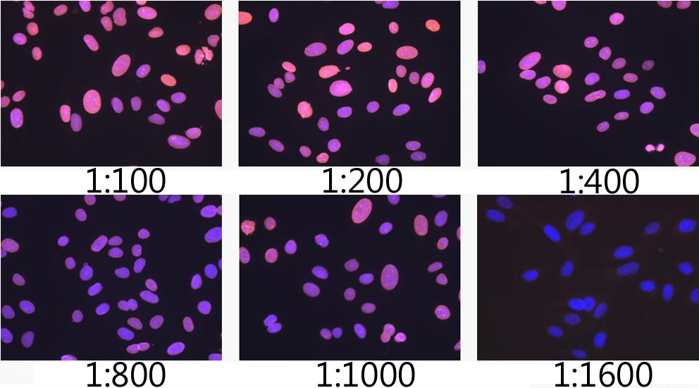 | Immunofluorescence analysis of U-2 OS cells using [KO Validated] Lamin A/C Rabbit mAb (A19524) at dilution of 1:100 - 1:1600. Blue: DAPI for nuclear staining. |
 | Immunohistochemistry analysis of paraffin-embedded Human colon carcinoma tissue using [KO Validated] Lamin A/C Rabbit mAb (A19524) at a dilution of 1:1300 (40x lens). High pressure antigen retrieval performed with 0.01M Citrate Buffer (pH 6.0) prior to IHC staining. |
 | Immunohistochemistry analysis of paraffin-embedded Mouse intestin tissue using [KO Validated] Lamin A/C Rabbit mAb (A19524) at a dilution of 1:1300 (40x lens). High pressure antigen retrieval performed with 0.01M Citrate Buffer (pH 6.0) prior to IHC staining. |
You may also be interested in:

