Your shopping cart is empty!
")
KO-Validated SLC25A4/ANT1 Rabbit mAb (20 μl)
| Reactivity: | Human, Mouse, Rat |
| Applications: | WB, IF/IC, IP, ELISA |
| Host Species: | Rabbit |
| Isotype: | IgG |
| Clonality: | Monoclonal antibody |
| Gene Name: | solute carrier family 25 member 4 |
| Gene Symbol: | SLC25A4 |
| Synonyms: | T1; ANT; AAC1; ANT1; PEO2; PEO3; ANT 1; PEOA2; MTDPS12; MTDPS12A |
| Gene ID: | 291 |
| UniProt ID: | P12235 |
| Clone ID: | 3C1Q3 |
| Immunogen: | A synthetic peptide corresponding to a sequence within amino acids 100-200 of human SLC25A4/ANT1 (NP_001142.2). |
| Dilution: | WB 1:1000-1:2000; IF/IC 1:100-1:1000 |
| Purification Method: | Affinity purification |
| Concentration: | 1 mg/ml |
| Buffer: | PBS with 0.05% proclin300, 0.05% BSA, 50% glycerol, pH7.3. |
| Storage: | Store at -20°C. Avoid freeze / thaw cycles. |
| Documents: | Manual-SLC25A4 monoclonal antibody |
Background
The gene SLC25A4 is a member of the mitochondrial carrier subfamily of solute carrier protein genes. The product of this gene functions as a gated pore that translocates ADP from the cytoplasm into the mitochondrial matrix and ATP from the mitochondrial matrix into the cytoplasm. The protein forms a homodimer embedded in the inner mitochondria membrane. Mutations in this gene have been shown to result in autosomal dominant progressive external opthalmoplegia and familial hypertrophic cardiomyopathy.
Images
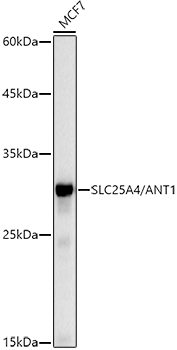 | Western blot analysis of lysates from MCF7 cells, using [KO Validated] SLC25A4/ANT1 Rabbit mAb (A20842) at1:1000 dilution. Secondary antibody: HRP-conjugated Goat anti-Rabbit IgG (H+L) (AS014) at1:10000 dilution. Lysates/proteins: 25μg per lane. Blocking buffer: 3% nonfat dry milk in TBST. Detection: ECL Basic Kit (RM00020). Exposure time: 10s. |
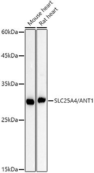 | Western blot analysis of various lysates using [KO Validated] SLC25A4/ANT1 Rabbit mAb (A20842) at1:1000 dilution. Secondary antibody: HRP-conjugated Goat anti-Rabbit IgG (H+L) (AS014) at1:10000 dilution. Lysates/proteins: 25μg per lane. Blocking buffer: 3% nonfat dry milk in TBST. Detection: ECL Basic Kit (RM00020). Exposure time: 60s. |
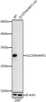 | Western blot analysis of lysates from wild type (WT) and SLC25A4/ANT1 knockout (KO) 293T cells, using [KO Validated] SLC25A4/ANT1 Rabbit mAb (A20842) at 1:1000 dilution. Secondary antibody: HRP-conjugated Goat anti-Rabbit IgG (H+L) (AS014) at 1:10000 dilution. Lysates/proteins: 25μg per lane. Blocking buffer: 3% nonfat dry milk in TBST. Detection: ECL Basic Kit (RM00020). Exposure time: 10s. |
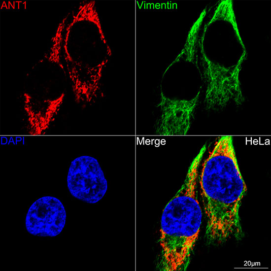 | Confocal imaging of HeLa cells using [KO Validated] SLC25A4/ANT1 Rabbit mAb (A20842, dilution 1:100) (Red). The cells were counterstained with [KO Validated] Vimentin Rabbit mAb (A19607, dilution 1:100) (Green). DAPI was used for nuclear staining (blue). Objective: 60x. |
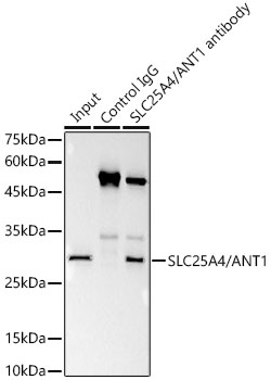 | Immunoprecipitation analysis of 300 μg extracts of 293T cells using 3 μg SLC25A4/ANT1 antibody (A20842). Western blot was performed from the immunoprecipitate using SLC25A4/ANT1 antibody (A20842) at a dilution of 1:1000. |
You may also be interested in:

