Your shopping cart is empty!
")
| Reactivity: | Human, Mouse, Rat |
| Applications: | WB, IHC, IF/IC, ELISA |
| Host Species: | Rabbit |
| Isotype: | IgG |
| Clonality: | Polyclonal antibody |
| Gene Name: | mitogen-activated protein kinase 3 |
| Gene Symbol: | MAPK3 |
| Synonyms: | ERK1; ERT2; ERK-1; PRKM3; P44ERK1; P44MAPK; HS44KDAP; HUMKER1A; p44-ERK1; p44-MAPK; K1 |
| Gene ID: | 5595 |
| UniProt ID: | P27361 |
| Immunogen: | Recombinant fusion protein containing a sequence corresponding to amino acids 1-100 of human ERK1 (NP_002737.2). |
| Dilution: | WB 1:500-1:2000; IHC 1:50-1:200; IF/IC 1:50-1:200 |
| Purification Method: | Affinity purification |
| Concentration: | 0.32 mg/ml |
| Buffer: | PBS with 0.02% sodium azide, 50% glycerol ,pH7.3. |
| Storage: | Store at -20°C. Avoid freeze / thaw cycles. |
| Documents: | Manual-MAPK3 polyclonal antibody |
Background
The protein encoded by this gene is a member of the MAP kinase family. MAP kinases, also known as extracellular signal-regulated kinases (ERKs), act in a signaling cascade that regulates various cellular processes such as proliferation, differentiation, and cell cycle progression in response to a variety of extracellular signals. This kinase is activated by upstream kinases, resulting in its translocation to the nucleus where it phosphorylates nuclear targets. Alternatively spliced transcript variants encoding different protein isoforms have been described.
Images
 | Western blot analysis of various lysates using [KD Validated] ERK1 Rabbit pAb (A0228) at 1:1000 dilution. Secondary antibody: HRP-conjugated Goat anti-Rabbit IgG (H+L) (AS014) at 1:10000 dilution. Lysates/proteins: 25μg per lane. Blocking buffer: 3% nonfat dry milk in TBST. |
 | Western blot analysis of lysates from wild type (WT) and ERK1 knockdown (KD) 293T cells, using [KD Validated] ERK1 Rabbit pAb (A0228) at 1:1000 dilution. Secondary antibody: HRP-conjugated Goat anti-Rabbit IgG (H+L) (AS014) at 1:10000 dilution. Lysates/proteins: 25μg per lane. Blocking buffer: 3% nonfat dry milk in TBST. Detection: ECL Basic Kit (RM00020). Exposure time: 10s. |
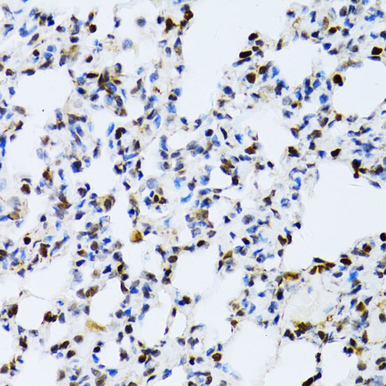 | Immunohistochemistry analysis of paraffin-embedded Rat lung using [KD Validated] ERK1 Rabbit pAb (A0228) at dilution of 1:100 (40x lens). Microwave antigen retrieval performed with 0.01M PBS Buffer (pH 7.2) prior to IHC staining. |
 | Immunohistochemistry analysis of paraffin-embedded Human kidney using [KD Validated] ERK1 Rabbit pAb (A0228) at dilution of 1:100 (40x lens). Microwave antigen retrieval performed with 0.01M PBS Buffer (pH 7.2) prior to IHC staining. |
 | Immunohistochemistry analysis of paraffin-embedded Mouse heart using [KD Validated] ERK1 Rabbit pAb (A0228) at dilution of 1:100 (40x lens). Microwave antigen retrieval performed with 0.01M PBS Buffer (pH 7.2) prior to IHC staining. |
You may also be interested in:

