Your shopping cart is empty!
")
| Reactivity: | Human, Mouse, Rat |
| Applications: | WB, IP, ELISA |
| Host Species: | Rabbit |
| Isotype: | IgG |
| Clonality: | Monoclonal antibody |
| Gene Name: | mitogen-activated protein kinase 8 |
| Gene Symbol: | MAPK8 |
| Synonyms: | JNK; [KD Validated] JNK1 |
| Gene ID: | 5599 |
| UniProt ID: | P45983 |
| Clone ID: | 6M4R7 |
| Immunogen: | A synthetic peptide corresponding to a sequence within amino acids 250-350 of human JNK1 (P45983). |
| Dilution: | WB 1:500-1:1000 |
| Purification Method: | Affinity purification |
| Concentration: | 0.6 mg/mL |
| Buffer: | PBS with 0.02% sodium azide, 0.05% BSA, 50% glycerol, pH7.3. |
| Storage: | Store at -20°C. Avoid freeze / thaw cycles. |
| Documents: | Manual-MAPK8 monoclonal antibody |
Background
The protein encoded by this gene is a member of the MAP kinase family. MAP kinases act as an integration point for multiple biochemical signals, and are involved in a wide variety of cellular processes such as proliferation, differentiation, transcription regulation and development. This kinase is activated by various cell stimuli, and targets specific transcription factors, and thus mediates immediate-early gene expression in response to cell stimuli. The activation of this kinase by tumor-necrosis factor alpha (TNF-alpha) is found to be required for TNF-alpha induced apoptosis. This kinase is also involved in UV radiation induced apoptosis, which is thought to be related to cytochrom c-mediated cell death pathway. Studies of the mouse counterpart of this gene suggested that this kinase play a key role in T cell proliferation, apoptosis and differentiation. Several alternatively spliced transcript variants encoding distinct isoforms have been reported.
Images
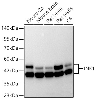 | Western blot analysis of various lysates using [KD Validated] JNK1 Rabbit mAb (A21888) at 1:1000 dilution. Secondary antibody: HRP-conjugated Goat anti-Rabbit IgG (H+L) (AS014) at 1:10000 dilution. Lysates/proteins: 25μg per lane. Blocking buffer: 3% nonfat dry milk in TBST. Detection: ECL Basic Kit (RM00020). Exposure time: 180s. |
 | Western blot analysis of lysates from wild type(WT) and JNK1 knockdown (KD) 293T cells, using [KD Validated] JNK1 Rabbit mAb (A21888) at 1:1000 dilution. Secondary antibody: HRP-conjugated Goat anti-Rabbit IgG (H+L) (AS014) at 1:10000 dilution. Lysates/proteins: 25μg per lane. Blocking buffer: 3% nonfat dry milk in TBST. Detection: ECL Basic Kit (RM00020). Exposure time: 180s. |
 | Immunoprecipitation of [KD Validated] JNK1 from 500 µg extracts of C6 cells was performed using 2 µg of [KD Validated] JNK1 Rabbit mAb (A21888). Rabbit IgG isotype control (AC042) was used to precipitate the Control IgG sample. IP samples were eluted with 1X non-reducing Laemmli Buffer. The Input lane represents 10% of the total input. Western blot analysis of immunoprecipitates was conducted using [KD Validated] JNK1 Rabbit mAb (A21888) at a dilution of 1:1000. |
You may also be interested in:

