Your shopping cart is empty!
| Reactivity: | Human |
| Applications: | WB, IF/ICC,IP, ELISA |
| Host Species: | Rabbit |
| Isotype: | IgG |
| Clonality: | Monoclonal antibody |
| Gene Name: | methylcrotonyl-CoA carboxylase subunit 1 |
| Gene Symbol: | MCCC1 |
| Synonyms: | MCCA; MCC-B; MCCCalpha; [KD Validated] MCCC1 |
| Gene ID: | 56922 |
| UniProt ID: | Q96RQ3 |
| Clone ID: | 4B4B7 |
| Immunogen: | Recombinant Protein corresponding to a sequence within amino acids 640-725 of human MCCC1(NP_064551.3). |
| Dilution: | WB 1:500-1:2000; IF/IC 1:50-1:200 |
| Purification Method: | Affinity purification |
| Concentration: | 1.75 mg/mL |
| Buffer: | PBS with 0.05% proclin300, 0.05% BSA, 50% glycerol, pH7.3. |
| Storage: | Store at -20°C. Avoid freeze / thaw cycles. |
| Documents: | Manual-MCCC1 monoclonal antibody |
Background
This gene encodes the large subunit of 3-methylcrotonyl-CoA carboxylase. This enzyme functions as a heterodimer and catalyzes the carboxylation of 3-methylcrotonyl-CoA to form 3-methylglutaconyl-CoA. Mutations in this gene are associated with 3-Methylcrotonylglycinuria, an autosomal recessive disorder of leucine catabolism.
Images
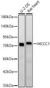 | Western blot analysis of various lysates using [KD Validated] MCCC1 Rabbit mAb (A25002) at 1:3000 dilution. Secondary antibody: HRP-conjugated Goat anti-Rabbit IgG (H+L) (AS014) at 1:10000 dilution. Lysates/proteins: 25μg per lane. Blocking buffer: 3% nonfat dry milk in TBST. Detection: ECL Basic Kit (RM00020). Exposure time: 45s. |
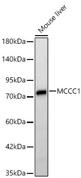 | Western blot analysis of lysates from Mouse liver using [KD Validated] MCCC1 Rabbit mAb (A25002) at 1:3000 dilution. Secondary antibody: HRP-conjugated Goat anti-Rabbit IgG (H+L) (AS014) at 1:10000 dilution. Lysates/proteins: 25μg per lane. Blocking buffer: 3% nonfat dry milk in TBST. Detection: ECL Basic Kit (RM00020). Exposure time: 15s. |
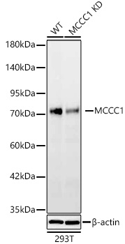 | Western blot analysis of lysates from wild type (WT) and MCCC1 knockdown (KD) 293T cells using [KD Validated] MCCC1 Rabbit mAb (A25002) at 1:3000 dilution. Secondary antibody: HRP-conjugated Goat anti-Rabbit IgG (H+L) (AS014) at 1:10000 dilution. Lysates/proteins: 25ug per lane. Blocking buffer: 3% nonfat dry milk in TBST. Detection: ECL Basic Kit (RM00020). Exposure time: 45s. |
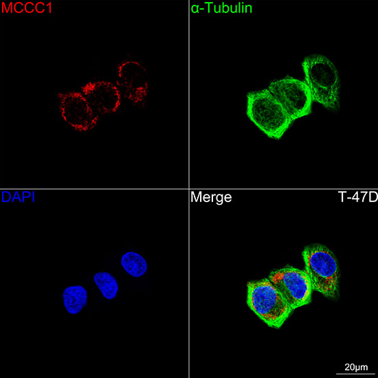 | Confocal imaging of T-47D cells using [KD Validated] MCCC1 Rabbit mAb (A25002,dilution 1:200) followed by a further incubation with Cy3 Goat Anti-Rabbit IgG (H+L) (AS007,dilution 1:500)(Red).The cells were counterstained with α-Tubulin Mouse mAb (AC012, dilution 1:400) followed by incubation with ABflo® 488-conjugated Goat Anti-Mouse IgG (H+L) Ab (AS076, dilution 1:500) (Green).DAPI was used for nuclear staining (Blue). Objective: 100x. |
 | Confocal imaging of T-47D cells using [KD Validated] MCCC1 Rabbit mAb (A25002,dilution 1:200) followed by a further incubation with Cy3 Goat Anti-Rabbit IgG (H+L) (AS007,dilution 1:500)(Red).The cells were counterstained with α-Tubulin Mouse mAb (AC012, dilution 1:400) followed by incubation with ABflo® 488-conjugated Goat Anti-Mouse IgG (H+L) Ab (AS076, dilution 1:500) (Green).DAPI was used for nuclear staining (Blue). Objective: 100x. |
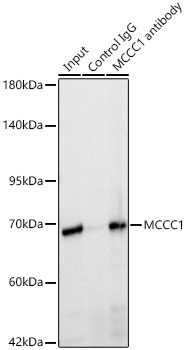 | Immunoprecipitation of MCCC1 in 300 µg extracts from T-47D cells using 3 µg [KD Validated] MCCC1 Rabbit mAb (A25002). Western blot analysis was performed using [KD Validated] MCCC1 Rabbit mAb (A25002) at 1:2000 dilution. |
You may also be interested in:

