Your shopping cart is empty!
KD-Validated MRPL20 Rabbit mAb (20 μl)
| Reactivity: | Human |
| Applications: | WB, IHC-P, ELISA |
| Host Species: | Rabbit |
| Isotype: | IgG |
| Clonality: | Monoclonal antibody |
| Gene Name: | mitochondrial ribosomal protein L20 |
| Gene Symbol: | MRPL20 |
| Synonyms: | L20mt; bL20m; MRP-L20 |
| Gene ID: | 55052 |
| UniProt ID: | Q9BYC9 |
| Immunogen: | A synthetic peptide corresponding to a sequence within amino acids 48-147 of human MRPL20 (NP_060441.2). |
| Dilution: | WB 1:1000-1:5000; IF/IC 1:50-1:200 |
| Purification Method: | Affinity purification |
| Concentration: | 1.33 mg/ml |
| Buffer: | PBS with 0.09% Sodium azide, 0.05% BSA, 50% glycerol, pH7.3. |
| Storage: | Store at -20°C. Avoid freeze / thaw cycles. |
| Documents: | Manual-MRPL20 monoclonal antibody |
Background
Mammalian mitochondrial ribosomal proteins are encoded by nuclear genes and help in protein synthesis within the mitochondrion. Mitochondrial ribosomes (mitoribosomes) consist of a small 28S subunit and a large 39S subunit. They have an estimated 75% protein to rRNA composition compared to prokaryotic ribosomes, where this ratio is reversed. Another difference between mammalian mitoribosomes and prokaryotic ribosomes is that the latter contain a 5S rRNA. Among different species, the proteins comprising the mitoribosome differ greatly in sequence, and sometimes in biochemical properties, which prevents easy recognition by sequence homology. This gene encodes a 39S subunit protein. A pseudogene corresponding to this gene is found on chromosome 21q. Alternative splicing results in multiple transcript variants encoding different isoforms.
Images
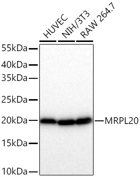 | Western blot analysis of various lysates using [KD Validated] MRPL20 Rabbit mAb (A26199) at 1:2000 dilution incubated overnight at 4℃. Secondary antibody: HRP-conjugated Goat anti-Rabbit IgG (H+L) (AS014) at 1:10000 dilution. Lysates/proteins: 25 μg per lane. Blocking buffer: 3% nonfat dry milk in TBST. Detection: ECL Basic Kit (RM00020). Exposure time: 10s. |
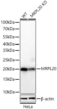 | Western blot analysis of lysates from wild type (WT) and MRPL20 knockdown (KD) HeLa cells using [KD Validated] MRPL20 Rabbit mAb (A26199) at 1:2000 dilution incubated overnight at 4℃. Secondary antibody: HRP-conjugated Goat anti-Rabbit IgG (H+L) (AS014) at 1:10000 dilution. Lysates/proteins: 25 μg per lane. Blocking buffer: 3% nonfat dry milk in TBST. Detection: ECL Basic Kit (RM00020). Exposure time: 1s. |
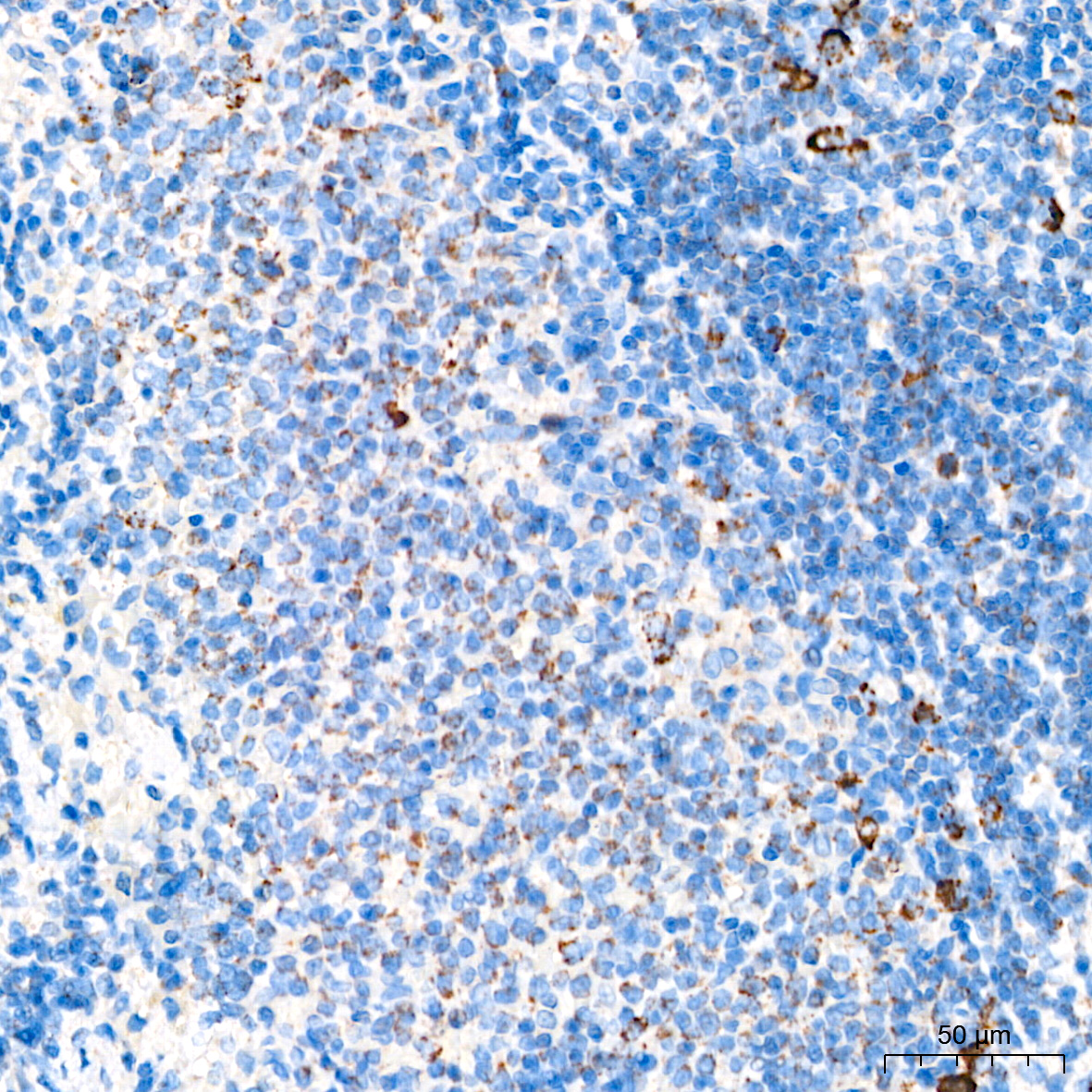 | Immunohistochemistry analysis of paraffin-embedded Rat spleen tissue using [KD Validated] MRPL20 Rabbit mAb (A26199) at a dilution of 1:200 (40x lens). High pressure antigen retrieval performed with 0.01M Citrate Buffer(pH 6.0) prior to IHC staining. |
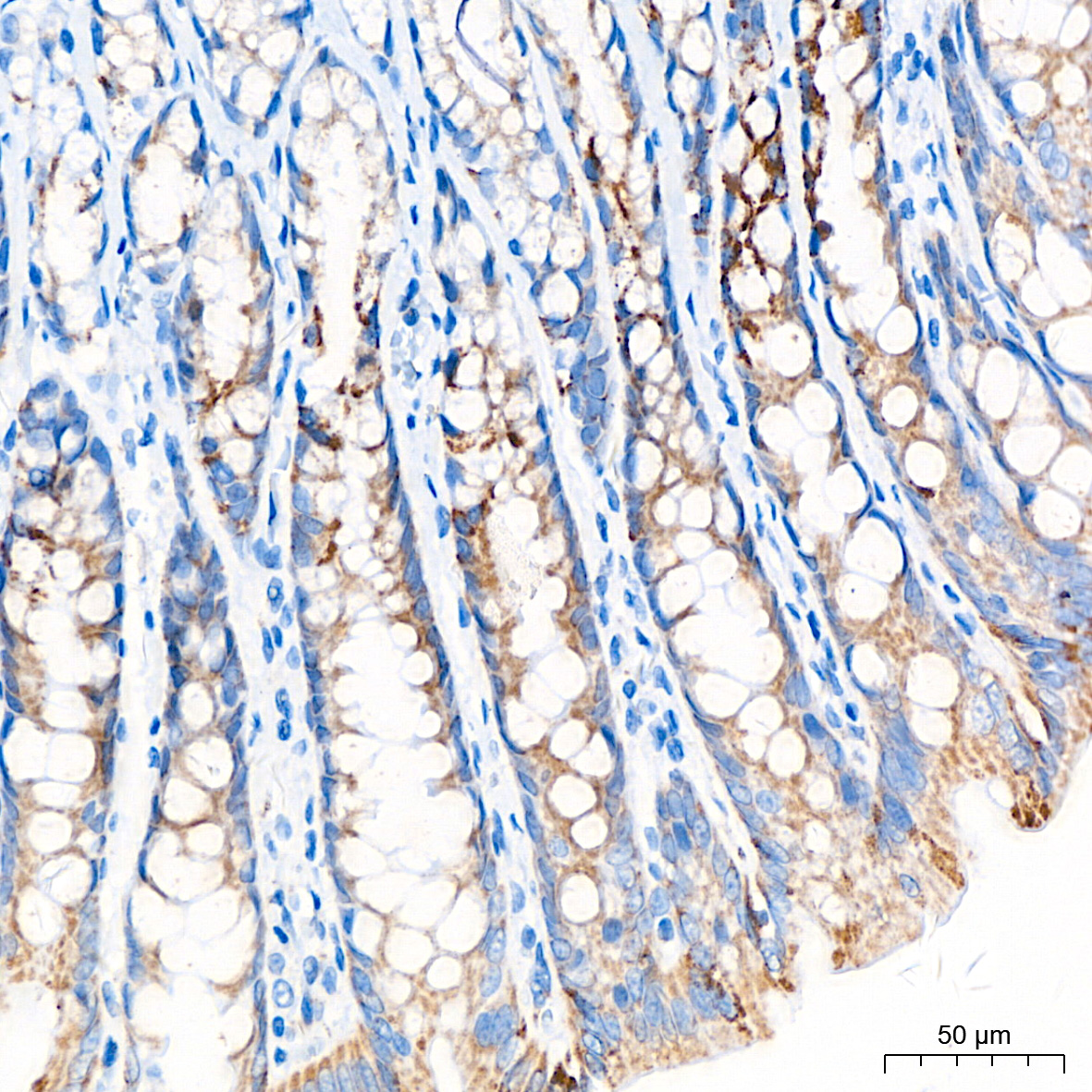 | Immunohistochemistry analysis of paraffin-embedded Rat colon tissue using [KD Validated] MRPL20 Rabbit mAb (A26199) at a dilution of 1:200 (40x lens). High pressure antigen retrieval performed with 0.01M Citrate Buffer(pH 6.0) prior to IHC staining. |
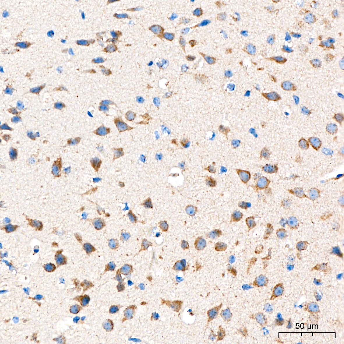 | Immunohistochemistry analysis of paraffin-embedded Mouse brain tissue using [KD Validated] MRPL20 Rabbit mAb (A26199) at a dilution of 1:200 (40x lens). High pressure antigen retrieval performed with 0.01M Citrate Buffer(pH 6.0) prior to IHC staining. |
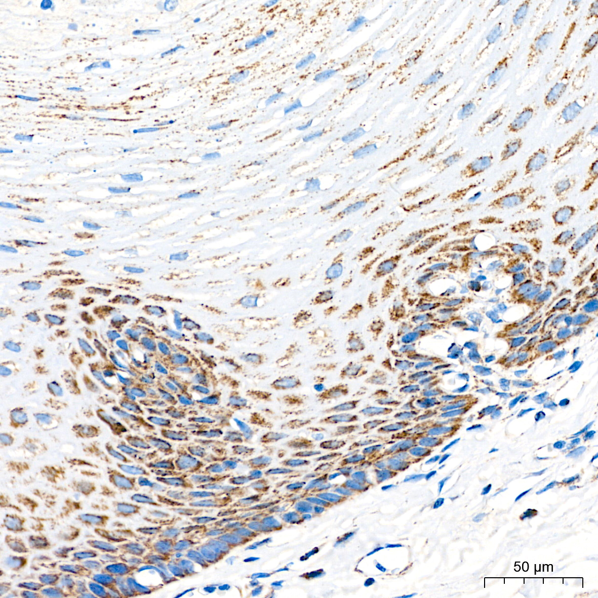 | Immunohistochemistry analysis of paraffin-embedded Human esophagus tissue using [KD Validated] MRPL20 Rabbit mAb (A26199) at a dilution of 1:200 (40x lens). High pressure antigen retrieval performed with 0.01M Citrate Buffer(pH 6.0) prior to IHC staining. |
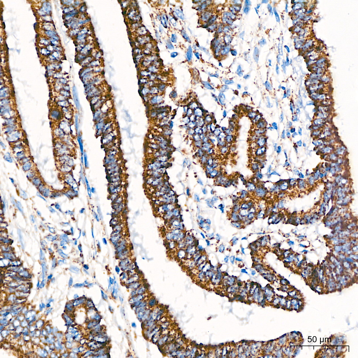 | Immunohistochemistry analysis of paraffin-embedded Human colon carcinoma tissue using [KD Validated] MRPL20 Rabbit mAb (A26199) at a dilution of 1:200 (40x lens). High pressure antigen retrieval performed with 0.01M Citrate Buffer(pH 6.0) prior to IHC staining. |
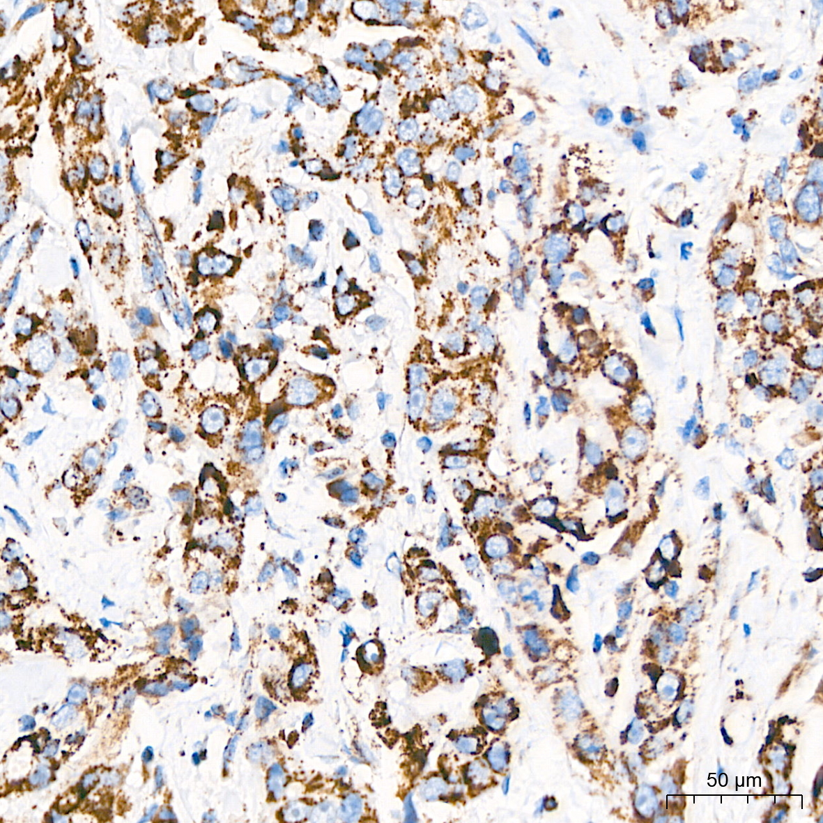 | Immunohistochemistry analysis of paraffin-embedded Human breast cancer tissue using [KD Validated] MRPL20 Rabbit mAb (A26199) at a dilution of 1:200 (40x lens). High pressure antigen retrieval performed with 0.01M Citrate Buffer(pH 6.0) prior to IHC staining. |
You may also be interested in:

