Your shopping cart is empty!
| Reactivity: | Human |
| Applications: | WB, ELISA |
| Host Species: | Rabbit |
| Isotype: | IgG |
| Clonality: | Polyclonal antibody |
| Gene Name: | mitochondrial ribosomal protein S7 |
| Gene Symbol: | MRPS7 |
| Synonyms: | MRPS7; MRP-S; MRP-S7; RP-S7; RPMS7; S7mt; bMRP27a; mitochondrial ribosomal protein S7 |
| Gene ID: | 51081 |
| UniProt ID: | Q9Y2R9 |
| Immunogen: | A synthetic peptide corresponding to a sequence within amino acids 40-140 of human MRPS7 (NP_057055.2). |
| Dilution: | WB 1:1000-1:5000; IHC 1:50-1:200 |
| Purification Method: | Affinity purification |
| Concentration: | 1.03 mg/mL |
| Buffer: | PBS with 0.05% proclin300, 50% glycerol, pH7.3. |
| Storage: | Store at -20°C. Avoid freeze / thaw cycles. |
| Documents: | Manual-MRPS7 polyclonal antibody |
Background
Mammalian mitochondrial ribosomal proteins are encoded by nuclear genes and help in protein synthesis within the mitochondrion. Mitochondrial ribosomes (mitoribosomes) consist of a small 28S subunit and a large 39S subunit. They have an estimated 75% protein to rRNA composition compared to prokaryotic ribosomes, where this ratio is reversed. Another difference between mammalian mitoribosomes and prokaryotic ribosomes is that the latter contain a 5S rRNA. Among different species, the proteins comprising the mitoribosome differ greatly in sequence, and sometimes in biochemical properties, which prevents easy recognition by sequence homology. This gene encodes a 28S subunit protein. In the prokaryotic ribosome, the comparable protein is thought to play an essential role in organizing the 3' domain of the 16 S rRNA in the vicinity of the P- and A-sites. Pseudogenes corresponding to this gene are found on chromosomes 8p and 12p.
Images
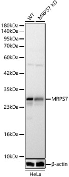 | Western blot analysis of lysates from wild type (WT) and MRPS7 knockdown (KD) HeLa cells using [KD Validated] MRPS7 Rabbit pAb (A24774) at 1:1000 dilution. Secondary antibody: HRP-conjugated Goat anti-Rabbit IgG (H+L) (AS014) at 1:10000 dilution.Lysates/proteins: 25 μg per lane. Blocking buffer: 3% nonfat dry milk in TBST. Detection: ECL Basic Kit (RM00020). Exposure time: 5s. |
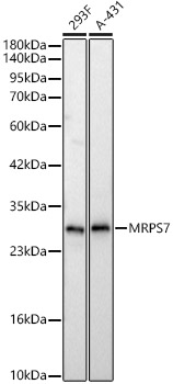 | Western blot analysis of various lysates using [KD Validated] MRPS7 Rabbit pAb (A24774) at 1:1000 dilution. Secondary antibody: HRP-conjugated Goat anti-Rabbit IgG (H+L) (AS014) at 1:10000 dilution. Lysates / proteins: 25 μg per lane. Blocking buffer: 3 % nonfat dry milk in TBST. Detection: ECL Basic Kit (RM00020). Exposure time: 5s. |
You may also be interested in:

