Your shopping cart is empty!
")
| Reactivity: | Human, Mouse, Rat |
| Applications: | WB, IF/IC, IP, ELISA |
| Host Species: | Rabbit |
| Isotype: | IgG |
| Clonality: | Polyclonal antibody |
| Gene Name: | NF2, moesin-ezrin-radixin like (MERLIN) tumor suppressor |
| Gene Symbol: | NF2 |
| Synonyms: | ACN; SCH; BANF; merlin-1; F2 |
| Gene ID: | 4771 |
| UniProt ID: | P35240 |
| Immunogen: | Recombinant fusion protein containing a sequence corresponding to amino acids 477-576 of human NF2 (NP_000259.1). |
| Dilution: | WB 1:500-1:2000; IF/IC 1:50-1:200 |
| Purification Method: | Affinity purification |
| Concentration: | 2.66 mg/ml |
| Buffer: | PBS with 0.02% sodium azide, 50% glycerol ,pH7.3. |
| Storage: | Store at -20°C. Avoid freeze / thaw cycles. |
| Documents: | Manual-NF2 polyclonal antibody |
Background
This gene encodes a protein that is similar to some members of the ERM (ezrin, radixin, moesin) family of proteins that link cytoskeletal components with proteins in the cell membrane. The encoded protein is involved in regulation of contact-dependent inhibition of cell proliferation and functions in cell-cell adhesion and transmembrane signaling. The encoded protein has been shown to interact with cell-surface proteins, proteins involved in cytoskeletal dynamics, and proteins involved in regulating ion transport. Disruption of this protein's function has been implicated in tumorigenesis and metastasis. Mutations in this gene are associated with neurofibromatosis type II which is characterized by nervous system and skin tumors and ocular abnormalities.
Images
 | Western blot analysis of lysates from HepG2 cells, using [KD Validated] NF2 Rabbit pAb (A0739) at 1:1000 dilution. Secondary antibody: HRP-conjugated Goat anti-Rabbit IgG (H+L) (AS014) at 1:10000 dilution. Lysates/proteins: 25μg per lane. Blocking buffer: 3% nonfat dry milk in TBST. Detection: ECL Basic Kit (RM00020). Exposure time: 3s. |
 | Western blot analysis of lysates from wild type (WT) and NF2 knockdown (KD) HeLa cells using [KD Validated] NF2 Rabbit pAb (A0739) at 1:1000 dilution incubated overnight at 4℃. Secondary antibody: HRP-conjugated Goat anti-Rabbit IgG (H+L) (AS014) at 1:10000 dilution. Lysates/proteins: 25 μg per lane. Blocking buffer: 3% nonfat dry milk in TBST. Detection: ECL Basic Kit (RM00020) Exposure time: 180 s. |
 | Western blot analysis of various lysates using [KD Validated] NF2 Rabbit pAb (A0739) at 1:1000 dilution incubated overnight at 4℃. Secondary antibody: HRP-conjugated Goat anti-Rabbit IgG (H+L) (AS014) at 1:10000 dilution. Lysates/proteins: 25 μg per lane. Blocking buffer: 3% nonfat dry milk in TBST. Detection: ECL Basic Kit (RM00020) Exposure time: 30 s. |
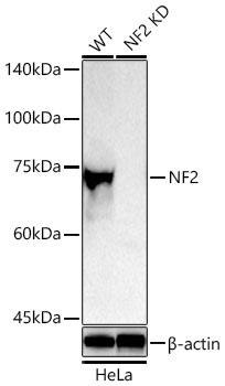
 | Western blot analysis of lysates from wild type (WT) and NF2 knockdown (KD) HeLa cells, using [KD Validated] NF2 Rabbit pAb (A0739) at 1:1000 dilution. Secondary antibody: HRP-conjugated Goat anti-Rabbit IgG (H+L) (AS014) at 1:10000 dilution. Lysates/proteins: 25μg per lane. Blocking buffer: 3% nonfat dry milk in TBST. Detection: ECL Basic Kit (RM00020). Exposure time: 3s. |
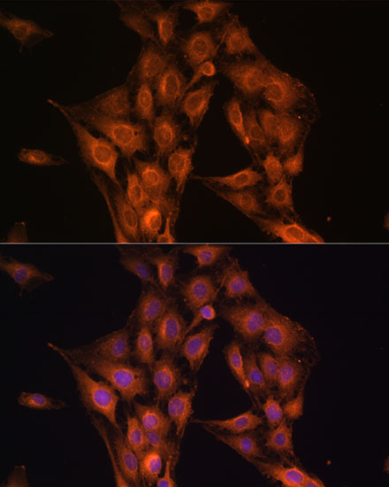
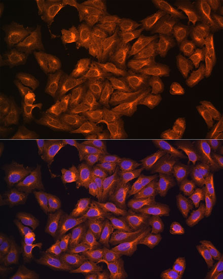 | Immunofluorescence analysis of C6 cells using [KD Validated] NF2 Rabbit pAb (A0739) at dilution of 1:100. Blue: DAPI for nuclear staining. |
 | Immunofluorescence analysis of L929 cells using [KD Validated] NF2 Rabbit pAb (A0739) at dilution of 1:100. Blue: DAPI for nuclear staining. |
 | Immunofluorescence analysis of U2OS cells using [KD Validated] NF2 Rabbit pAb (A0739) at dilution of 1:100. Blue: DAPI for nuclear staining. |
 | Immunoprecipitation analysis of 200 μg extracts of SKOV3 cells using 3 μg [KD Validated] NF2 Rabbit pAb (A0739). Western blot was performed from the immunoprecipitate using NF2 antibody (A0739) at a dilution of 1:1000. |
You may also be interested in:

