Your shopping cart is empty!
")
| Reactivity: | Human, Mouse, Rat |
| Applications: | WB, IHC, IF/IC, ELISA |
| Host Species: | Rabbit |
| Isotype: | IgG |
| Clonality: | Monoclonal antibody |
| Gene Name: | ornithine aminotransferase |
| Gene Symbol: | OAT |
| Synonyms: | OKT; GACR; HOGA; OATASE |
| Gene ID: | 4942 |
| UniProt ID: | P04181 |
| Immunogen: | Recombinant fusion protein containing a sequence corresponding to amino acids 160-439 of human OAT (NP_000265.1). |
| Dilution: | WB 1:11000-1:44000; IHC 1:1000-1:10000; IF/IC 1:200-1:800 |
| Purification Method: | Affinity purification |
| Concentration: | 1.18 mg/ml |
| Buffer: | PBS with 0.09% Sodium azide, 0.05% BSA, 50% glycerol, pH7.3. |
| Storage: | Store at -20°C. Avoid freeze / thaw cycles. |
| Documents: | Manual-OAT monoclonal antibody |
Background
This gene encodes the mitochondrial enzyme ornithine aminotransferase, which is a key enzyme in the pathway that converts arginine and ornithine into the major excitatory and inhibitory neurotransmitters glutamate and GABA. Mutations that result in a deficiency of this enzyme cause the autosomal recessive eye disease Gyrate Atrophy. Alternatively spliced transcript variants encoding different isoforms have been described. Related pseudogenes have been defined on the X chromosome.
Images
 | Western blot analysis of various lysates using [KD Validated] OAT Rabbit mAb (A27191) at 1:11000 dilution incubated at room temperature for 1.5 hours. Secondary antibody: HRP-conjugated Goat anti-Rabbit IgG (H+L) (AS014) at 1:10000 dilution. Lysates/proteins: 25 μg per lane. Blocking buffer: 3% nonfat dry milk in TBST. Detection: ECL Basic Kit (RM00020). Exposure time: 20s. |
 | Western blot analysis of lysates from wild type (WT) and OAT knockdown (KD) 293T cells using [KD Validated] OAT Rabbit mAb (A27191) at 1:11000 dilution incubated at room temperature for 1.5 hours. Secondary antibody: HRP-conjugated Goat anti-Rabbit IgG (H+L) (AS014) at 1:10000 dilution. Lysates/proteins: 25 μg per lane. Blocking buffer: 3% nonfat dry milk in TBST. Detection: ECL Basic Kit (RM00020). Exposure time: 20s. |
 | Immunohistochemistry analysis of paraffin-embedded Human liver tissue using [KD Validated] OAT Rabbit mAb (A27191) at a dilution of 1:6000 (40x lens). High pressure antigen retrieval performed with 0.01M Tris-EDTA Buffer (pH 9.0) prior to IHC staining. |
 | Immunohistochemistry analysis of paraffin-embedded Human kidney tissue using [KD Validated] OAT Rabbit mAb (A27191) at a dilution of 1:6000 (40x lens). High pressure antigen retrieval performed with 0.01M Tris-EDTA Buffer (pH 9.0) prior to IHC staining. |
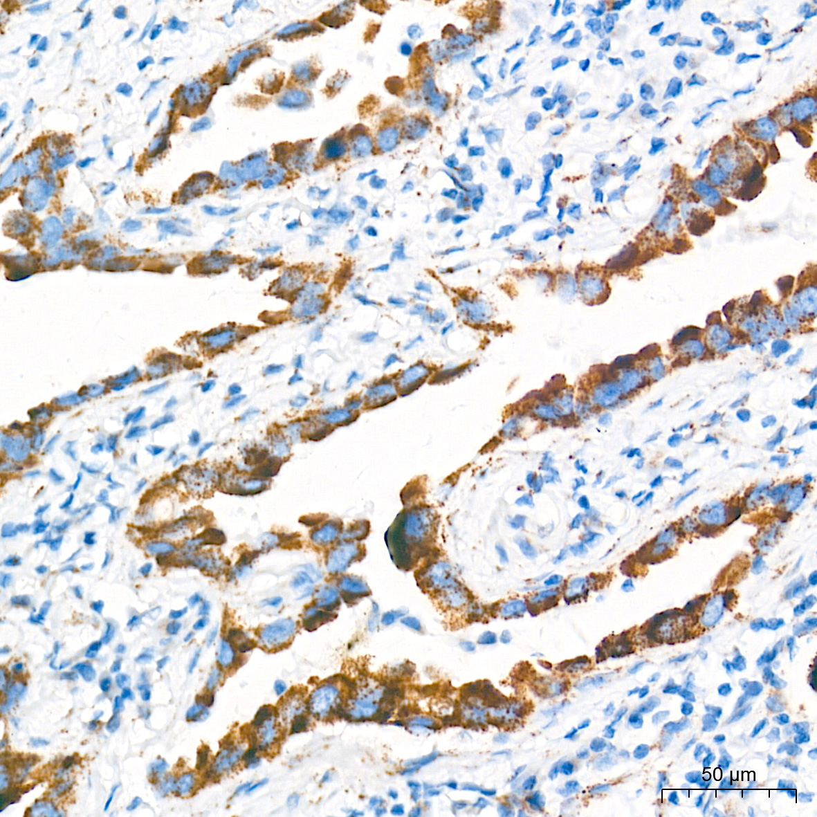 | Immunohistochemistry analysis of paraffin-embedded Human lung cancer tissue using [KD Validated] OAT Rabbit mAb (A27191) at a dilution of 1:6000 (40x lens). High pressure antigen retrieval performed with 0.01M Tris-EDTA Buffer (pH 9.0) prior to IHC staining. |
 | Immunohistochemistry analysis of paraffin-embedded Mouse liver tissue using [KD Validated] OAT Rabbit mAb (A27191) at a dilution of 1:6000 (40x lens). High pressure antigen retrieval performed with 0.01M Tris-EDTA Buffer (pH 9.0) prior to IHC staining. |
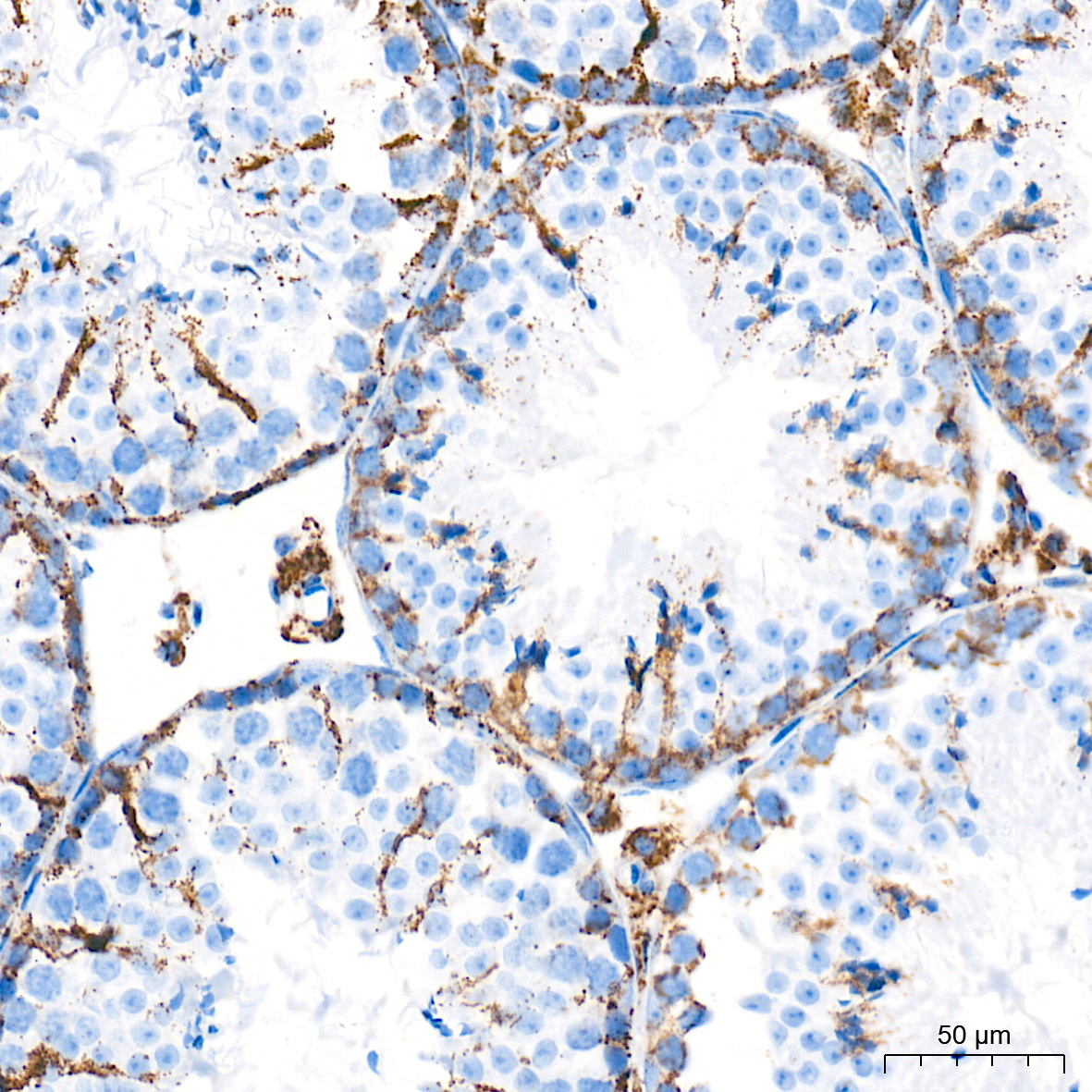 | Immunohistochemistry analysis of paraffin-embedded Mouse testis tissue using [KD Validated] OAT Rabbit mAb (A27191) at a dilution of 1:6000 (40x lens). High pressure antigen retrieval performed with 0.01M Tris-EDTA Buffer (pH 9.0) prior to IHC staining. |
 | Immunohistochemistry analysis of paraffin-embedded Rat colon tissue using [KD Validated] OAT Rabbit mAb (A27191) at a dilution of 1:6000 (40x lens). High pressure antigen retrieval performed with 0.01M Tris-EDTA Buffer (pH 9.0) prior to IHC staining. |
You may also be interested in:

