Your shopping cart is empty!
")
| Reactivity: | Human |
| Applications: | WB, IHC, ELISA |
| Host Species: | Rabbit |
| Isotype: | IgG |
| Clonality: | Monoclonal antibody |
| Gene Name: | prosaposin |
| Gene Symbol: | PSAP |
| Synonyms: | GLBA; SAP1; SAP2; PSAPD; PARK24; [KD Validated] PSAP |
| Gene ID: | 5660 |
| UniProt ID: | P07602 |
| Clone ID: | 3G4E8 |
| Immunogen: | Recombinant fusion protein containing a sequence corresponding to amino acids 275-524 of human PSAP (NP_002769.1). |
| Dilution: | WB 1:500-1:1000; IHC 1:100-1:500 |
| Purification Method: | Affinity purification |
| Concentration: | 0.66 mg/mL |
| Buffer: | PBS with 0.05% proclin300, 0.05% BSA, 50% glycerol, pH7.3. |
| Storage: | Store at -20°C. Avoid freeze / thaw cycles. |
| Documents: | Manual-PSAP monoclonal antibody |
Background
This gene encodes a highly conserved preproprotein that is proteolytically processed to generate four main cleavage products including saposins A, B, C, and D. Each domain of the precursor protein is approximately 80 amino acid residues long with nearly identical placement of cysteine residues and glycosylation sites. Saposins A-D localize primarily to the lysosomal compartment where they facilitate the catabolism of glycosphingolipids with short oligosaccharide groups. The precursor protein exists both as a secretory protein and as an integral membrane protein and has neurotrophic activities. Mutations in this gene have been associated with Gaucher disease and metachromatic leukodystrophy. Alternative splicing results in multiple transcript variants, at least one of which encodes an isoform that is proteolytically processed.
Images
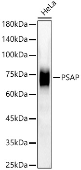 | Western blot analysis of lysates from HeLa cells using [KD Validated] PSAP Rabbit mAb (A22736) at 1:1000 dilution incubated overnight at 4℃. Secondary antibody: HRP-conjugated Goat anti-Rabbit IgG (H+L) (AS014) at 1:10000 dilution. Lysates/proteins: 25 μg per lane. Blocking buffer: 3% nonfat dry milk in TBST. Detection: ECL Enhanced Kit (RM00021). Exposure time: 60s. |
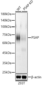 | Western blot analysis of lysates from wild type(WT) and PSAP knockdown (KD) 293T cells using [KD Validated] PSAP Rabbit mAb (A22736) at 1:1000 dilution incubated overnight at 4℃. Secondary antibody: HRP-conjugated Goat anti-Rabbit IgG (H+L) (AS014) at 1:10000 dilution. Lysates/proteins: 25 μg per lane. Blocking buffer: 3% nonfat dry milk in TBST. Detection: ECL Enhanced Kit (RM00021). Exposure time: 60s. |
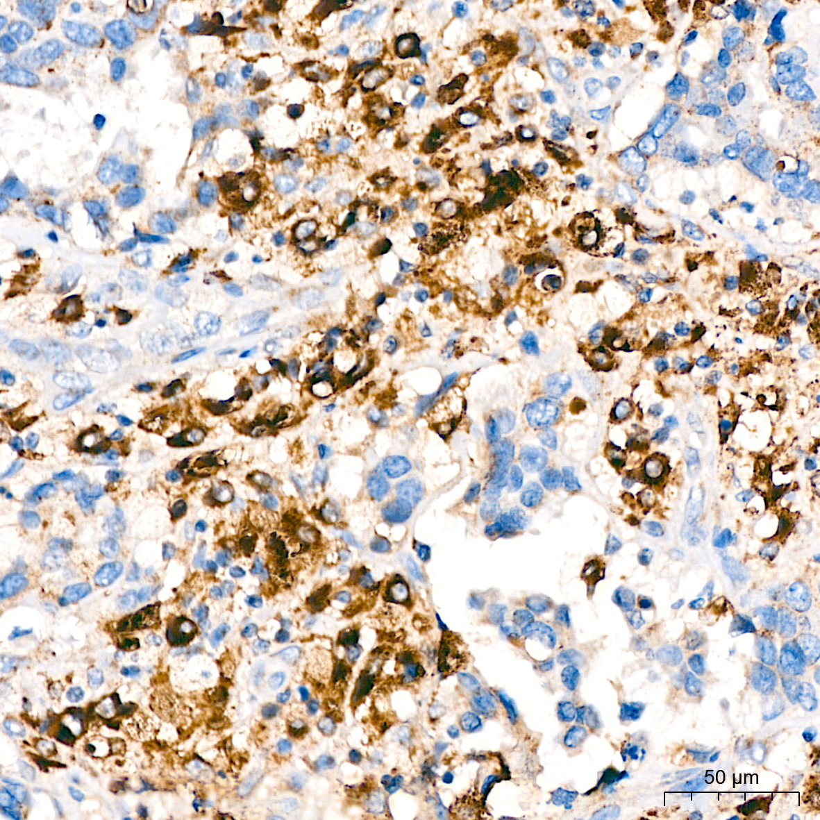 | Immunohistochemistry analysis of paraffin-embedded Human breast cancer tissue using [KD Validated] PSAP Rabbit mAb (A22736) at a dilution of 1:400 (40x lens). High pressure antigen retrieval performed with 0.01M Citrate Bufferr (pH 6.0) prior to IHC staining. |
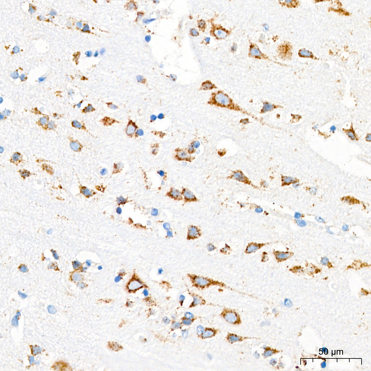 | Immunohistochemistry analysis of paraffin-embedded Human brain tissue using [KD Validated] PSAP Rabbit mAb (A22736) at a dilution of 1:400 (40x lens). High pressure antigen retrieval performed with 0.01M Citrate Bufferr (pH 6.0) prior to IHC staining. |
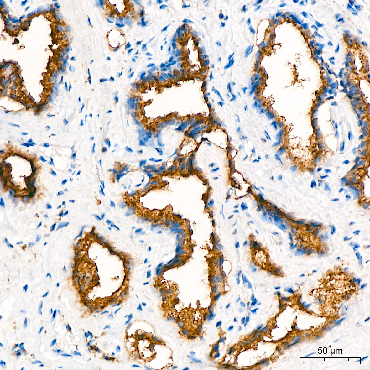 | Immunohistochemistry analysis of paraffin-embedded Human prostate cancer tissue using [KD Validated] PSAP Rabbit mAb (A22736) at a dilution of 1:400 (40x lens). High pressure antigen retrieval performed with 0.01M Citrate Bufferr (pH 6.0) prior to IHC staining. |
You may also be interested in:

