Your shopping cart is empty!
| Reactivity: | Human |
| Applications: | WB, IHC-P, ELISA |
| Host Species: | Rabbit |
| Isotype: | IgG |
| Clonality: | Monoclonal antibody |
| Gene Name: | Rac GTPase activating protein 1 |
| Gene Symbol: | RACGAP1 |
| Synonyms: | CYK4; CDAN3B; ID-GAP; HsCYK-4; MgcRacGAP; [KD Validated] RGAP1 |
| Gene ID: | 29127 |
| UniProt ID: | Q9H0H5 |
| Clone ID: | 3B5F3 |
| Immunogen: | A synthetic peptide corresponding to a sequence within amino acids 533-632 of human RGAP1 (NP_037409.2). |
| Dilution: | WB 1:1000-1:5000 |
| Purification Method: | Affinity purification |
| Concentration: | 0.247 mg/mL |
| Buffer: | PBS with 0.05% proclin300, 0.05% BSA, 50% glycerol, pH7.3. |
| Storage: | Store at -20°C. Avoid freeze / thaw cycles. |
| Documents: | Manual-RACGAP1 monoclonal antibody |
Background
This gene encodes a GTPase-activating protein (GAP) that is a compoment of the centralspindlin complex. This protein binds activated forms of Rho GTPases and stimulates GTP hydrolysis, which results in negative regulation of Rho-mediated signals. This protein plays a regulatory role in cytokinesis, cell growth, and differentiation. Alternatively spliced transcript variants have been found for this gene. There is a pseudogene for this gene on chromosome 12.
Images
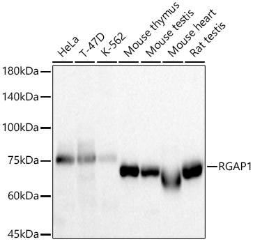 | Western blot analysis of various lysates, using [KD Validated] RGAP1 Rabbit mAb (A24948) at 1:2000 dilution. Secondary antibody: HRP-conjugated Goat anti-Rabbit IgG (H+L) (AS014) at 1:10000 dilution. Lysates/proteins: 25μg per lane. Blocking buffer: 3% nonfat dry milk in TBST. Detection: ECL Basic Kit (RM00020). Exposure time: 10s. |
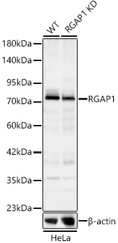 | Western blot analysis of lysates from wild type (WT) and RGAP1 knockdown (KD) HeLa cells using [KD Validated] RGAP1 Rabbit mAb (A24948) at 1:800 dilution. Secondary antibody: HRP-conjugated Goat anti-Rabbit IgG (H+L) (AS014) at 1:10000 dilution. Lysates/proteins: 25ug per lane. Blocking buffer: 3% nonfat dry milk in TBST. Detection: ECL Basic Kit (RM00020). Exposure time: 30s. |
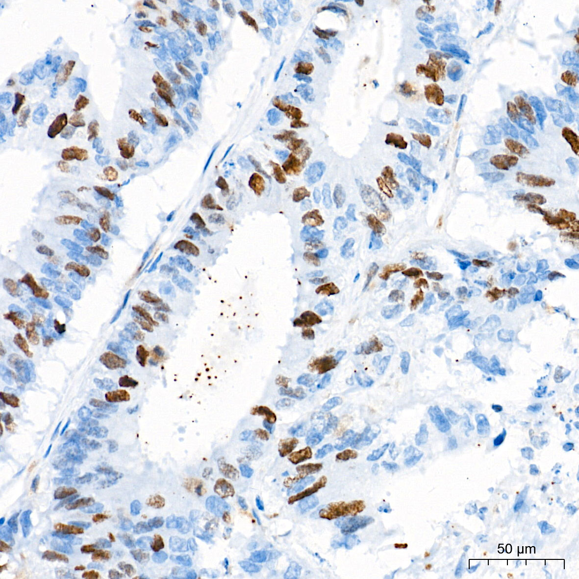 | Immunohistochemistry analysis of paraffin-embedded Human colon carcinoma tissue using [KD Validated] RGAP1 Rabbit mAb (A24948) at a dilution of 1:200 (40x lens). High pressure antigen retrieval performed with 0.01M Tris-EDTA Buffer (pH 9.0) prior to IHC staining. |
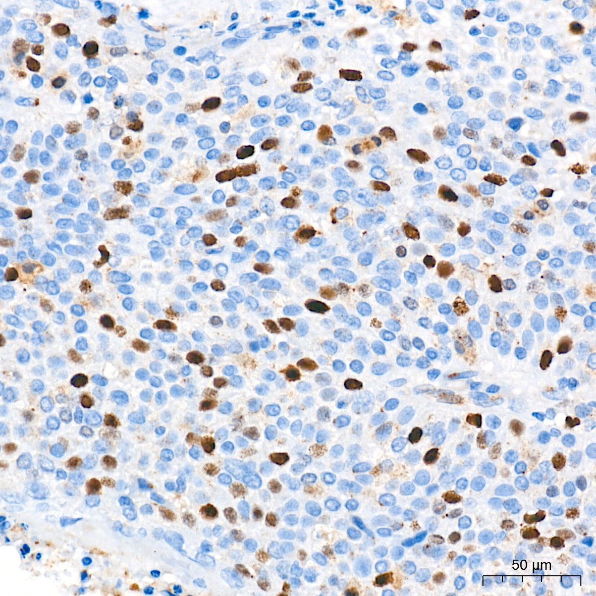 | Immunohistochemistry analysis of paraffin-embedded Human cervix cancer tissue using [KD Validated] RGAP1 Rabbit mAb (A24948) at a dilution of 1:200 (40x lens). High pressure antigen retrieval performed with 0.01M Tris-EDTA Buffer (pH 9.0) prior to IHC staining. |
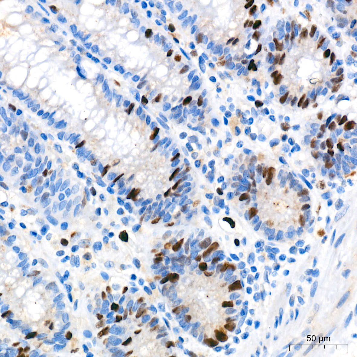 | Immunohistochemistry analysis of paraffin-embedded Human colon tissue using [KD Validated] RGAP1 Rabbit mAb (A24948) at a dilution of 1:200 (40x lens). High pressure antigen retrieval performed with 0.01M Tris-EDTA Buffer (pH 9.0) prior to IHC staining. |
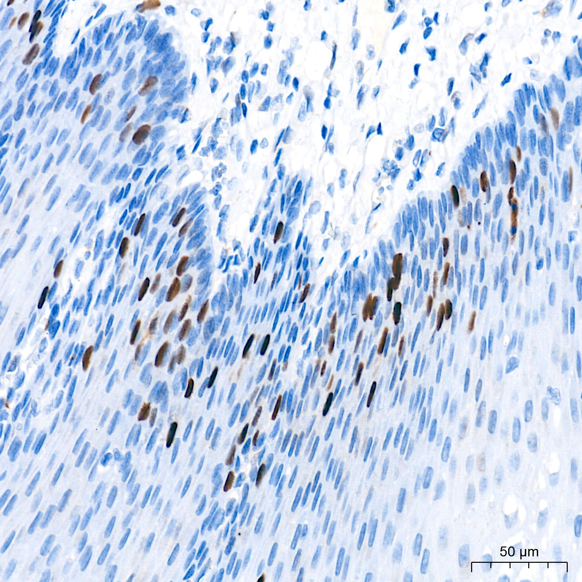 | Immunohistochemistry analysis of paraffin-embedded Human esophagus tissue using [KD Validated] RGAP1 Rabbit mAb (A24948) at a dilution of 1:200 (40x lens). High pressure antigen retrieval performed with 0.01M Tris-EDTA Buffer (pH 9.0) prior to IHC staining. |
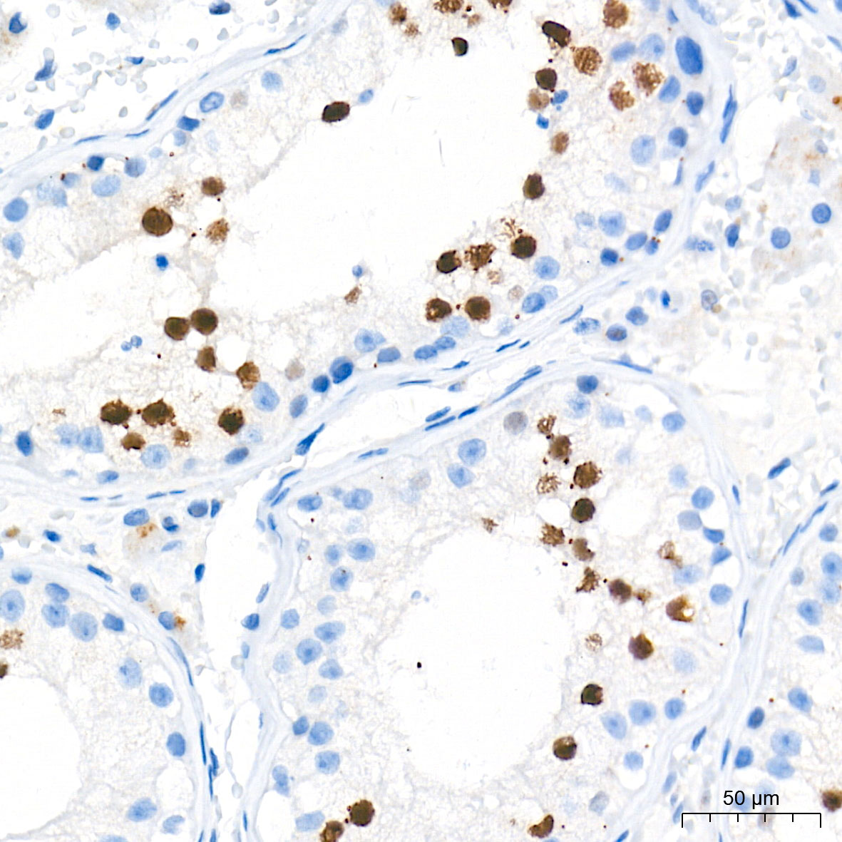 | Immunohistochemistry analysis of paraffin-embedded Human testis tissue using [KD Validated] RGAP1 Rabbit mAb (A24948) at a dilution of 1:200 (40x lens). High pressure antigen retrieval performed with 0.01M Tris-EDTA Buffer (pH 9.0) prior to IHC staining. |
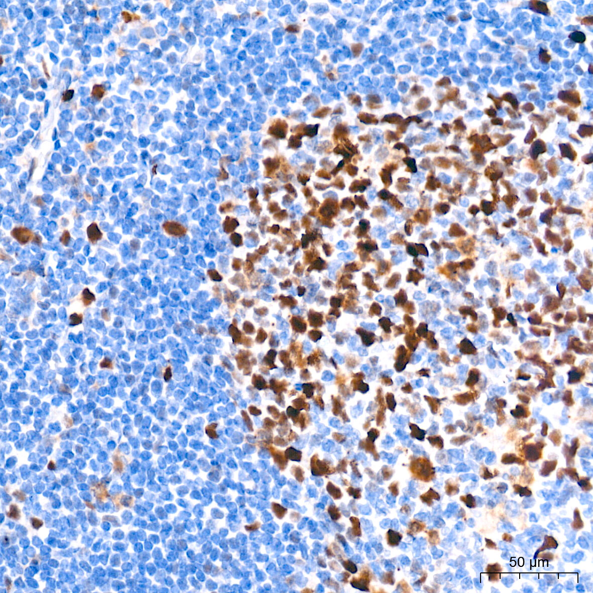 | Immunohistochemistry analysis of paraffin-embedded Human tonsil tissue using [KD Validated] RGAP1 Rabbit mAb (A24948) at a dilution of 1:200 (40x lens). High pressure antigen retrieval performed with 0.01M Tris-EDTA Buffer (pH 9.0) prior to IHC staining. |
You may also be interested in:

