Your shopping cart is empty!
| Reactivity: | Human |
| Applications: | WB, IHC-P, ELISA |
| Host Species: | Rabbit |
| Isotype: | IgG |
| Clonality: | Monoclonal antibody |
| Gene Name: | reactive oxygen species modulator 1 |
| Gene Symbol: | ROMO1 |
| Synonyms: | MTGM, MTGMP, C20orf52, bA353C18.2, ROMO1 |
| Gene ID: | 140823 |
| UniProt ID: | P60602 |
| Immunogen: | A synthetic peptide corresponding to a sequence within amino acids 1-79 of human ROMO1(NP_542786.1). |
| Purification Method: | Affinity purification |
| Concentration: | 1.45 mg/ml |
| Buffer: | PBS with 0.09% Sodium azide, 0.05% BSA, 50% glycerol, pH7.3. |
| Storage: | Store at -20°C. Avoid freeze / thaw cycles. |
| Documents: | Manual-ROMO1 monoclonal antibody |
Background
The protein encoded by this gene is a mitochondrial membrane protein that is responsible for increasing the level of reactive oxygen species (ROS) in cells. The protein also has antimicrobial activity against a variety of bacteria by inducing bacterial membrane breakage.
Images
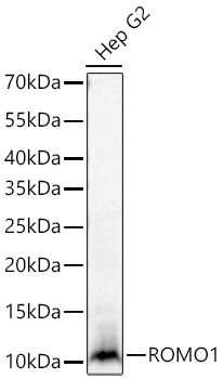 | Western blot analysis of lysates from Hep G2 cells using [KD Validated] ROMO1 Rabbit mAb (A27581) at 1:14000 dilution incubated at room temperature for 1.5 hours. Secondary antibody: HRP-conjugated Goat anti-Rabbit IgG (H+L) (AS014) at 1:10000 dilution. Lysates/proteins: 25 μg per lane. Blocking buffer: 3% nonfat dry milk in TBST. Detection: ECL Basic Kit (RM00020). Exposure time: 10s. |
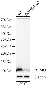 | Western blot analysis of lysates from wild type (WT) and ROMO1 knockdown (KD) 293T cells using [KD Validated] ROMO1 Rabbit mAb (A27581) at 1:14000 dilution incubated at room temperature for 1.5 hours. Secondary antibody: HRP-conjugated Goat anti-Rabbit IgG (H+L) (AS014) at 1:10000 dilution. Lysates/proteins: 25 μg per lane. Blocking buffer: 3% nonfat dry milk in TBST. Detection: ECL Basic Kit (RM00020). Exposure time: 20s. |
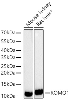 | Western blot analysis of various lysates using [KD Validated] ROMO1 Rabbit mAb (A27581) at 1:14000 dilution incubated at room temperature for 1.5 hours. Secondary antibody: HRP-conjugated Goat anti-Rabbit IgG (H+L) (AS014) at 1:10000 dilution. Lysates/proteins: 25 μg per lane. Blocking buffer: 3% nonfat dry milk in TBST. Detection: ECL Basic Kit (RM00020). Exposure time: 45s. |
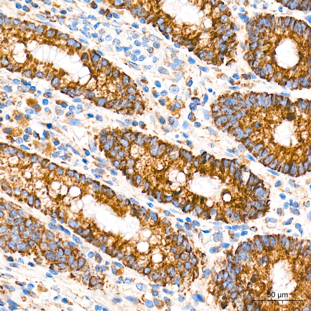 | Immunohistochemistry analysis of paraffin-embedded Human colon tissue using [KD Validated] ROMO1 Rabbit mAb (A27581) at a dilution of 1:700 (40x lens). High pressure antigen retrieval performed with 0.01M Tris-EDTA Buffer (pH 9.0) prior to IHC staining. |
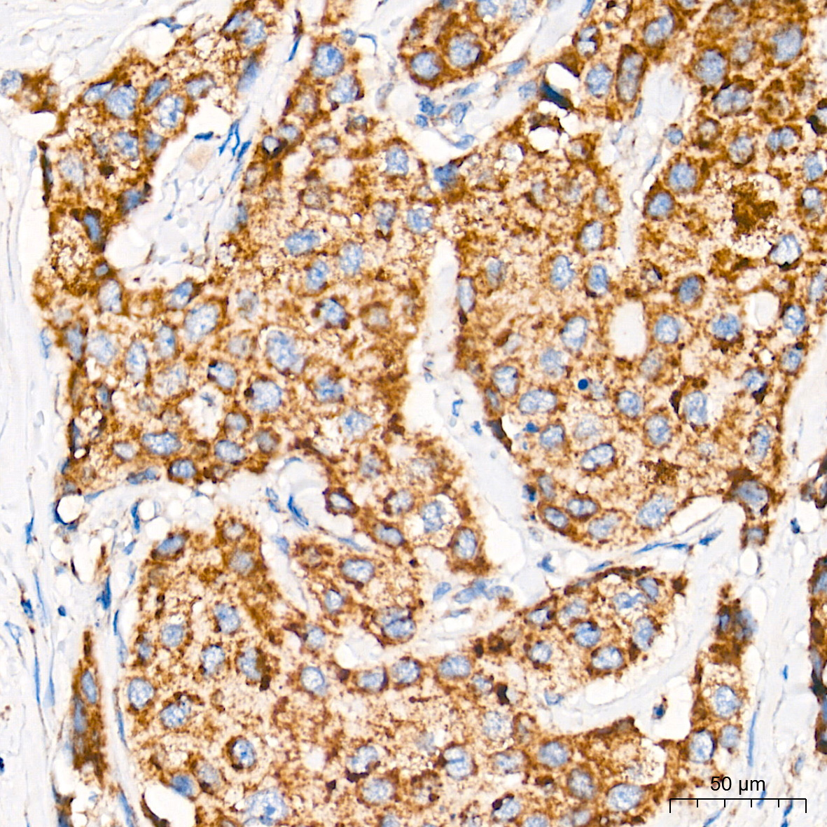 | Immunohistochemistry analysis of paraffin-embedded Human breast cancer tissue using [KD Validated] ROMO1 Rabbit mAb (A27581) at a dilution of 1:700 (40x lens). High pressure antigen retrieval performed with 0.01M Tris-EDTA Buffer (pH 9.0) prior to IHC staining. |
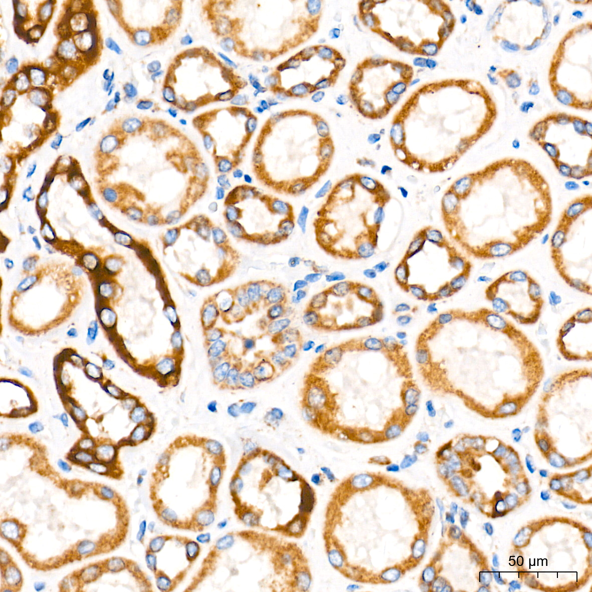 | Immunohistochemistry analysis of paraffin-embedded Human kidney tissue using [KD Validated] ROMO1 Rabbit mAb (A27581) at a dilution of 1:700 (40x lens). High pressure antigen retrieval performed with 0.01M Tris-EDTA Buffer (pH 9.0) prior to IHC staining. |
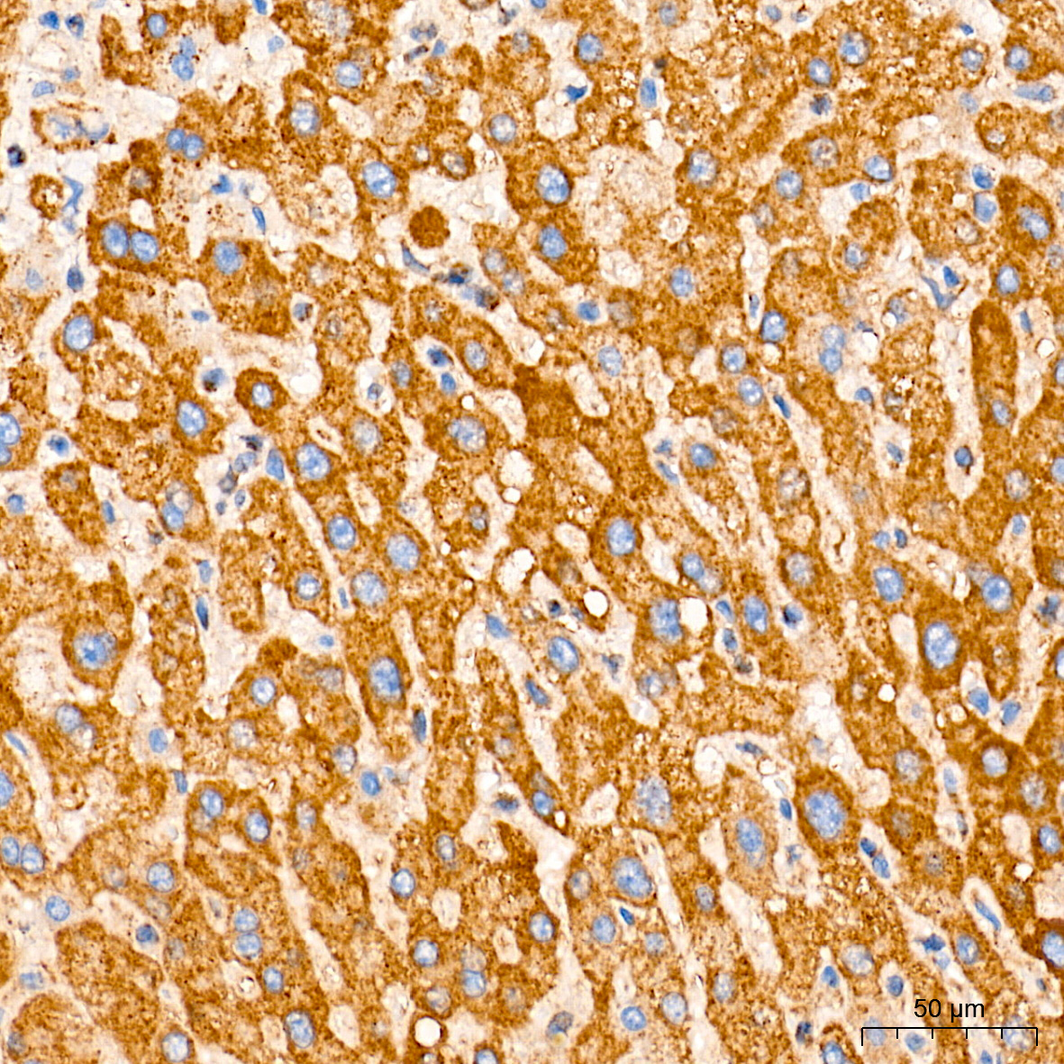 | Immunohistochemistry analysis of paraffin-embedded Human liver tissue using [KD Validated] ROMO1 Rabbit mAb (A27581) at a dilution of 1:700 (40x lens). High pressure antigen retrieval performed with 0.01M Tris-EDTA Buffer (pH 9.0) prior to IHC staining. |
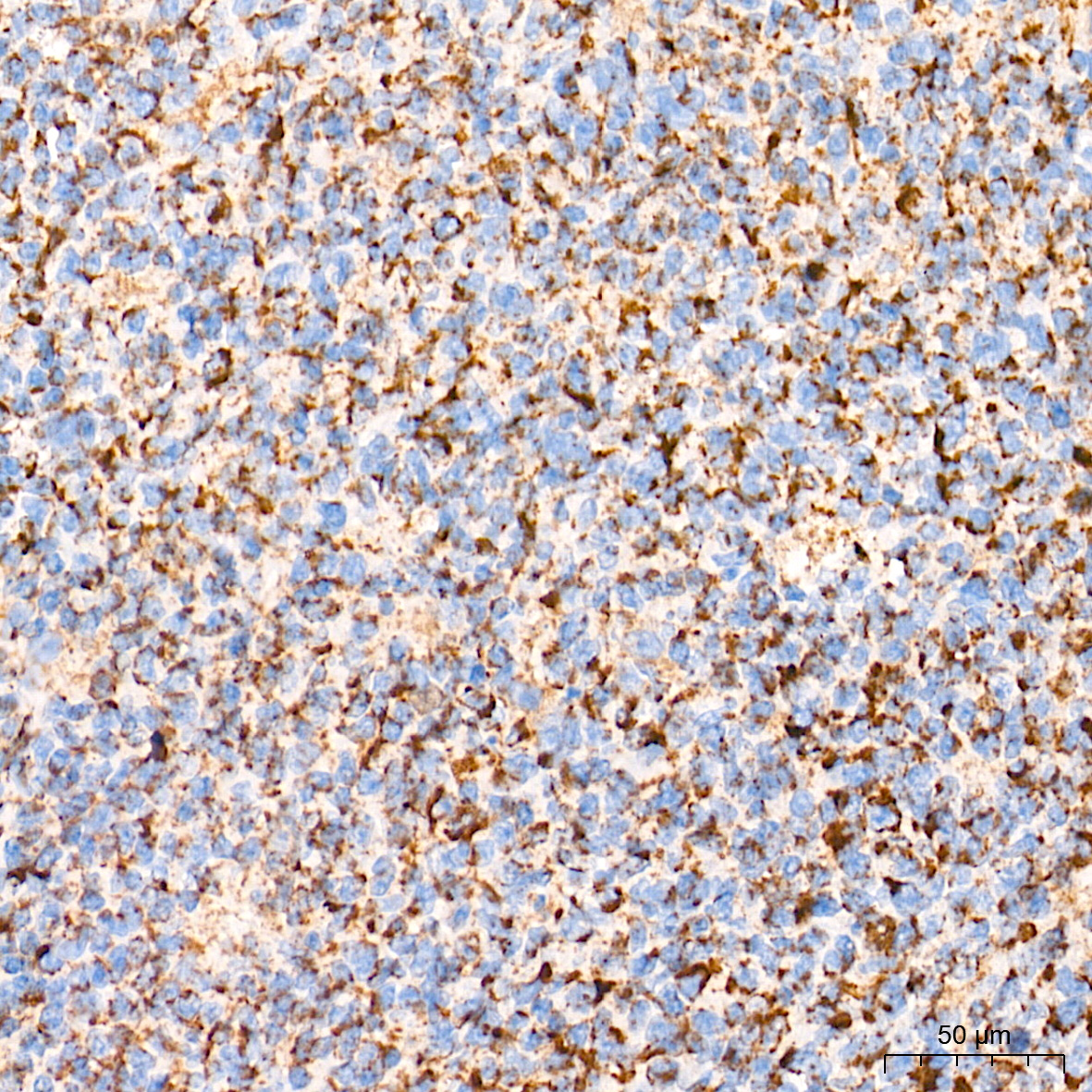 | Immunohistochemistry analysis of paraffin-embedded Human tonsil tissue using [KD Validated] ROMO1 Rabbit mAb (A27581) at a dilution of 1:700 (40x lens). High pressure antigen retrieval performed with 0.01M Tris-EDTA Buffer (pH 9.0) prior to IHC staining. |
You may also be interested in:

