Your shopping cart is empty!
KD-Validated Sharpin Rabbit mAb (20 μl)
| Reactivity: | Human |
| Applications: | WB, IF/ICC, ELISA |
| Host Species: | Rabbit |
| Isotype: | IgG |
| Clonality: | Monoclonal antibody |
| Gene Name: | SHANK associated RH domain interactor |
| Gene Symbol: | SHARPIN |
| Synonyms: | SIPL1; [KD Validated] Sharpin |
| Gene ID: | 81858 |
| UniProt ID: | Q9H0F6 |
| Clone ID: | 8B3S10 |
| Immunogen: | Recombinant Protein corresponding to a sequence within amino acids 1-126 of human SHARPIN(NP_112236.3). |
| Dilution: | WB 1:500-1:1000 |
| Purification Method: | Affinity purification |
| Concentration: | 1.61 mg/mL |
| Buffer: | PBS with 0.05% proclin300, 0.05% BSA, 50% glycerol, pH7.3. |
| Storage: | Store at -20°C. Avoid freeze / thaw cycles. |
| Documents: | Manual-SHARPIN monoclonal antibody |
Background
Enables polyubiquitin modification-dependent protein binding activity. Involved in protein linear polyubiquitination and regulation of signal transduction. Located in cytosol. Part of LUBAC complex.
Images
 | Western blot analysis of lysates from PC-3 cells, using [KD Validated] Sharpin Rabbit mAb (A24809) at 1:1000 dilution. Secondary antibody: HRP-conjugated Goat anti-Rabbit IgG (H+L) (AS014) at 1:10000 dilution. Lysates/proteins: 25μg per lane. Blocking buffer: 3% nonfat dry milk in TBST. Detection: ECL Basic Kit (RM00020). Exposure time: 30s. |
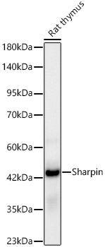 | Western blot analysis of lysates from Rat thymus, using [KD Validated] Sharpin Rabbit mAb (A24809) at 1:1000 dilution. Secondary antibody: HRP-conjugated Goat anti-Rabbit IgG (H+L) (AS014) at 1:10000 dilution. Lysates/proteins: 25μg per lane. Blocking buffer: 3% nonfat dry milk in TBST. Detection: ECL Basic Kit (RM00020). Exposure time: 90s. |
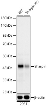 | Western blot analysis of lysates from wild type (WT) and Sharpin knockdown (KD) 293T cells, using [KD Validated] Sharpin Rabbit mAb (A24809) at 1:1000 dilution. Secondary antibody: HRP-conjugated Goat anti-Rabbit IgG (H+L) (AS014) at 1:10000 dilution. Lysates/proteins: 25ug per lane. Blocking buffer: 3% nonfat dry milk in TBST. Detection: ECL Basic Kit (RM00020). Exposure time: 30s. |
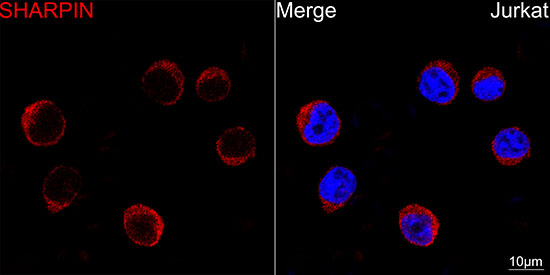 | Confocal imaging of Jurkat cells using [KD Validated] Sharpin Rabbit mAb (A24809,dilution 1:200) followed by a further incubation with Cy3 Goat Anti-Rabbit IgG (H+L) (AS007, dilution 1:500) (Red). DAPI was used for nuclear staining (blue). Objective: 100x. |
You may also be interested in:

