Your shopping cart is empty!
")
| Reactivity: | Human, Mouse, Rat |
| Applications: | WB, IF/IC, IP, ELISA |
| Host Species: | Rabbit |
| Isotype: | IgG |
| Clonality: | Monoclonal antibody |
| Gene Name: | SMAD family member 2 |
| Gene Symbol: | SMAD2 |
| Synonyms: | JV18; LDS6; CHTD8; MADH2; MADR2; JV18-1; hMAD-2; hSMAD2; [KD Validated] Smad2 |
| Gene ID: | 4087 |
| UniProt ID: | Q15796 |
| Clone ID: | 2E3D8 |
| Immunogen: | A synthetic peptide corresponding to a sequence within amino acids 1-100 of human Smad2 (Q15796). |
| Dilution: | WB 1:1000-1:2000; IF/IC 1:100-1:400 |
| Purification Method: | Affinity purification |
| Concentration: | 0.8 mg/mL |
| Buffer: | PBS with 0.02% sodium azide, 0.05% BSA, 50% glycerol, pH7.3. |
| Storage: | Store at -20°C. Avoid freeze / thaw cycles. |
| Documents: | Manual-SMAD2 monoclonal antibody |
Background
The protein encoded by this gene belongs to the SMAD, a family of proteins similar to the gene products of the Drosophila gene 'mothers against decapentaplegic' (Mad) and the C. elegans gene Sma. SMAD proteins are signal transducers and transcriptional modulators that mediate multiple signaling pathways. This protein mediates the signal of the transforming growth factor (TGF)-beta, and thus regulates multiple cellular processes, such as cell proliferation, apoptosis, and differentiation. This protein is recruited to the TGF-beta receptors through its interaction with the SMAD anchor for receptor activation (SARA) protein. In response to TGF-beta signal, this protein is phosphorylated by the TGF-beta receptors. The phosphorylation induces the dissociation of this protein with SARA and the association with the family member SMAD4. The association with SMAD4 is important for the translocation of this protein into the nucleus, where it binds to target promoters and forms a transcription repressor complex with other cofactors. This protein can also be phosphorylated by activin type 1 receptor kinase, and mediates the signal from the activin. Alternatively spliced transcript variants have been observed for this gene.
Images
 | Western blot analysis of lysates from wild type (WT) and Smad2 knockdown (KD) HeLa cells using [KD Validated] Smad2 Rabbit mAb (A19114) at 1:1000 dilution incubated overnight at 4℃. Secondary antibody: HRP-conjugated Goat anti-Rabbit IgG (H+L) (AS014) at 1:10000 dilution. Lysates/proteins: 25μg per lane. Blocking buffer: 3% nonfat dry milk in TBST. Detection: ECL Basic Kit (RM00020). Exposure time: 60s. |
 | Western blot analysis of various lysates using [KD Validated] Smad2 Rabbit mAb (A19114) at 1:1000 dilution incubated overnight at 4℃. Secondary antibody: HRP-conjugated Goat anti-Rabbit IgG (H+L) (AS014) at 1:10000 dilution. Lysates/proteins: 25μg per lane. Blocking buffer: 3% nonfat dry milk in TBST. Detection: ECL Basic Kit (RM00020). Exposure time: 90s. |
 | Western blot analysis of lysates from Rat lung using [KD Validated] Smad2 Rabbit mAb (A19114) at 1:1000 dilution incubated overnight at 4℃. Secondary antibody: HRP-conjugated Goat anti-Rabbit IgG (H+L) (AS014) at 1:10000 dilution. Lysates/proteins: 25μg per lane. Blocking buffer: 3% nonfat dry milk in TBST. Detection: ECL Basic Kit (RM00020). Exposure time: 3min. |
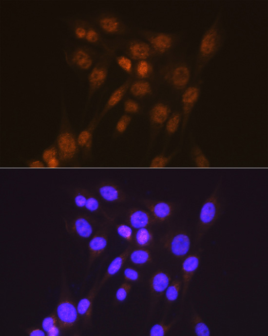
 | Immunofluorescence analysis of NIH/3T3 cells using [KD Validated] Smad2 Rabbit mAb (A19114) at a dilution of 1:100 (40x lens). Secondary antibody: Cy3 Goat anti-Rabbit IgG (H+L) (AS007) at 1:500 dilution. Blue: DAPI for nuclear staining. |
 | Confocal imaging of HeLa cells using [KD Validated] Smad2 Rabbit mAb (A19114, dilution 1:100) followed by a further incubation with Cy3 Goat Anti-Rabbit IgG (H+L) (AS007,dilution 1:500) (Red). The cells were counterstained with α-Tubulin Mouse mAb (AC012, dilution 1:400) followed by incubation with ABflo® 488-conjugated Goat Anti-Mouse IgG (H+L) Ab (AS076, dilution 1:500) (Green). DAPI was used for nuclear staining (Blue). Objective: 100x. |
 | Immunoprecipitation of Smad2 from 300 µg extracts of HeLa cells was performed using 3 µg of Smad2 antibody (A19114). Rabbit IgG isotype control (AC042) was used to precipitate the Control IgG sample. IP samples were eluted with 1X reducing Laemmli Buffer. The Input lane represents 10% of the total input. Western blot analysis of immunoprecipitates was conducted using Smad2 antibody (A19114) at a dilution of 1:1000. |
You may also be interested in:

