Your shopping cart is empty!
KD-Validated TIMM10 Rabbit mAb (20 μl)
| Reactivity: | Human |
| Applications: | WB, IHC, IF/IC, ELISA |
| Host Species: | Rabbit |
| Isotype: | IgG |
| Clonality: | Monoclonal antibody |
| Gene Name: | translocase of inner mitochondrial membrane 10 |
| Gene Symbol: | TIMM10 |
| Synonyms: | TIM10; TIM10A; TIMM10A; [KD Validated] TIMM10 |
| Gene ID: | 26519 |
| UniProt ID: | P62072 |
| Clone ID: | 9N5D1 |
| Immunogen: | Recombinant fusion protein containing a sequence corresponding to amino acids 1-90aa of human TIMM10 (NP_000204.3). |
| Dilution: | WB 1:2000-1:7000; IHC 1:500-1:1000; IF/IC 1:50-1:200 |
| Purification Method: | Affinity purification |
| Concentration: | 1.24 mg/mL |
| Buffer: | PBS with 0.05% proclin300, 0.05% BSA, 50% glycerol, pH7.3. |
| Storage: | Store at -20°C. Avoid freeze / thaw cycles. |
| Documents: | Manual-TIMM10 monoclonal antibody |
Background
The mitochondrial protein encoded by the gene TIMM10 belongs to a family of evolutionarily conserved proteins that are organized in heterooligomeric complexes in the mitochondrial intermembrane space. These proteins mediate the import and insertion of hydrophobic membrane proteins into the mitochondrial inner membrane, functioning as intermembrane space chaperones for the highly insoluble carrier proteins.
Images
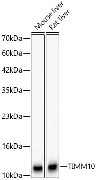 | Western blot analysis of various lysates using [KD Validated] TIMM10 Rabbit mAb (A24996) at 1:6000 dilution. Secondary antibody: HRP-conjugated Goat anti-Rabbit IgG (H+L) (AS014) at 1:10000 dilution. Lysates/proteins: 25μg per lane. Blocking buffer: 3% nonfat dry milk in TBST. Detection: ECL Basic Kit (RM00020). Exposure time: 30s. |
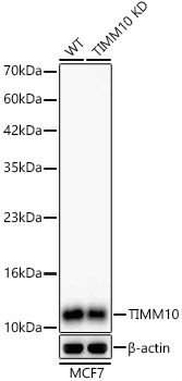 | Western blot analysis of lysates from wild type (WT) and TIMM10 knockdown (KD) MCF7 cells using [KD Validated] TIMM10 Rabbit mAb (A24996) at 1:6000 dilution. Secondary antibody: HRP-conjugated Goat anti-Rabbit IgG (H+L) (AS014) at 1:10000 dilution. Lysates/proteins: 25μg per lane. Blocking buffer: 3% nonfat dry milk in TBST. Detection: ECL Basic Kit (RM00020). Exposure time: 30s. |
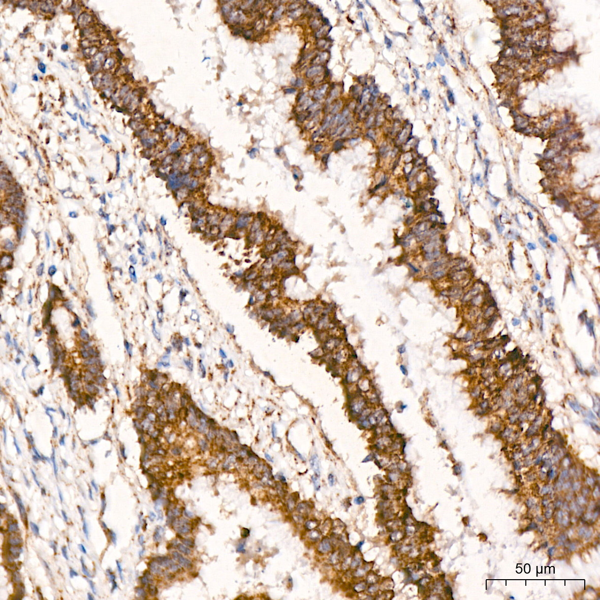 | Immunohistochemistry analysis of paraffin-embedded Human colon carcinoma tissue using [KD Validated] TIMM10 Rabbit mAb (A24996) at a dilution of 1:800 (40x lens). High pressure antigen retrieval performed with 0.01M Citrate Bufferr (pH 6.0) prior to IHC staining. |
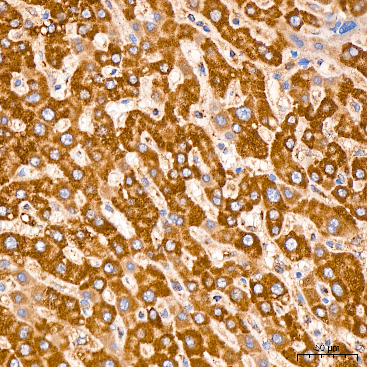 | Immunohistochemistry analysis of paraffin-embedded Human liver tissue using [KD Validated] TIMM10 Rabbit mAb (A24996) at a dilution of 1:800 (40x lens). High pressure antigen retrieval performed with 0.01M Citrate Bufferr (pH 6.0) prior to IHC staining. |
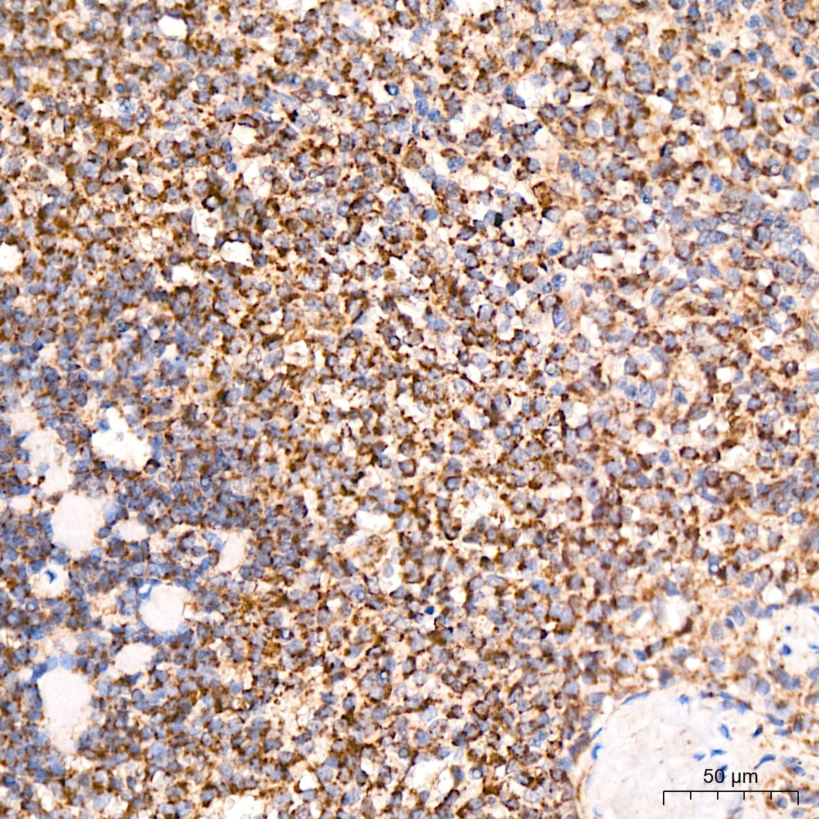 | Immunohistochemistry analysis of paraffin-embedded Human spleen tissue using [KD Validated] TIMM10 Rabbit mAb (A24996) at a dilution of 1:800 (40x lens). High pressure antigen retrieval performed with 0.01M Citrate Bufferr (pH 6.0) prior to IHC staining. |
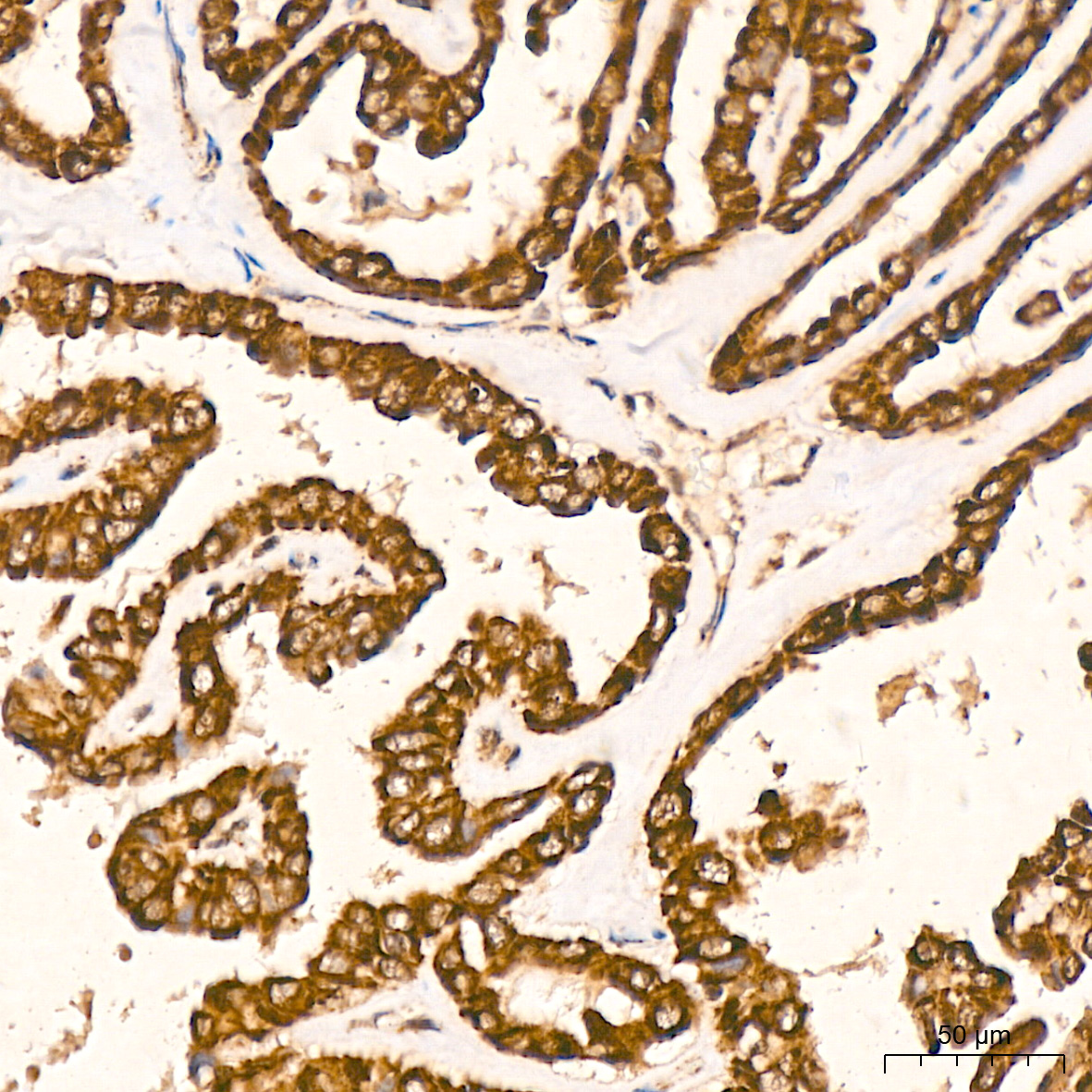 | Immunohistochemistry analysis of paraffin-embedded Human thyroid cancer tissue using [KD Validated] TIMM10 Rabbit mAb (A24996) at a dilution of 1:800 (40x lens). High pressure antigen retrieval performed with 0.01M Citrate Bufferr (pH 6.0) prior to IHC staining. |
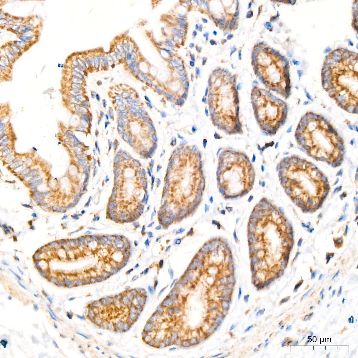 | Immunohistochemistry analysis of paraffin-embedded Rat colon tissue using [KD Validated] TIMM10 Rabbit mAb (A24996) at a dilution of 1:800 (40x lens). High pressure antigen retrieval performed with 0.01M Citrate Bufferr (pH 6.0) prior to IHC staining. |
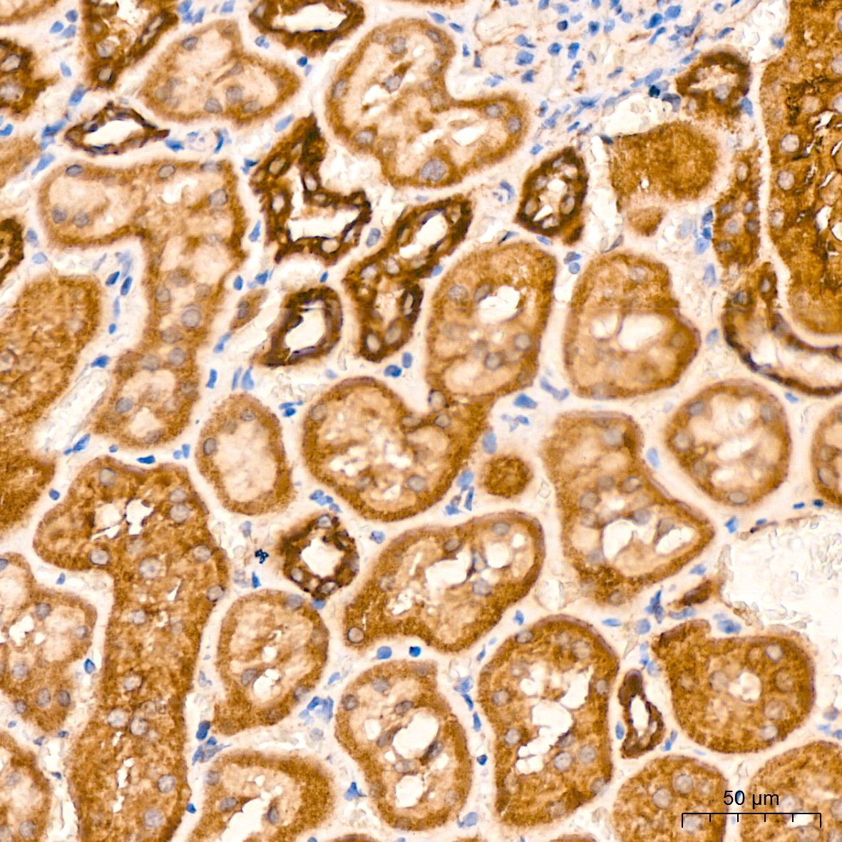 | Immunohistochemistry analysis of paraffin-embedded Rat kidney tissue using [KD Validated] TIMM10 Rabbit mAb (A24996) at a dilution of 1:800 (40x lens). High pressure antigen retrieval performed with 0.01M Citrate Bufferr (pH 6.0) prior to IHC staining. |
You may also be interested in:

