Your shopping cart is empty!
KD-Validated TIMM13 Rabbit mAb (20 μl)
| Reactivity: | Human |
| Applications: | WB, IF/IC, ELISA |
| Host Species: | Rabbit |
| Isotype: | IgG |
| Clonality: | Monoclonal antibody |
| Gene Name: | translocase of inner mitochondrial membrane 13 |
| Gene Symbol: | TIMM13 |
| Synonyms: | TWIST1; ACS3; BPES2; BPES3; CRS; CRS1; CSO; SCS; TWIST; bHLHa38; twist-related protein 1 |
| Gene ID: | 26517 |
| UniProt ID: | Q9Y5L4 |
| Clone ID: | 8O2U3 |
| Immunogen: | Recombinant fusion protein containing a sequence corresponding to amino acids 1-95 of human TIMM13 (NP_036590.1). |
| Dilution: | WB 1:500-1:1000; IF/IC 1:50-1:200 |
| Purification Method: | Affinity purification |
| Concentration: | 1.96 mg/mL |
| Buffer: | PBS with 0.05% proclin300, 0.05% BSA, 50% glycerol, pH7.3. |
| Storage: | Store at -20°C. Avoid freeze / thaw cycles. |
| Documents: | Manual-TIMM13 monoclonal antibody |
Background
The gene TIMM13 encodes a member of the evolutionarily conserved TIMM (translocase of inner mitochondrial membrane) family of proteins that function as chaperones in the import of proteins from the cytoplasm into the mitochondrial inner membrane. Proteins of this family play a role in collecting substrate proteins from the translocase of the outer mitochondrial membrane (TOM) complex and delivering them to either the sorting and assembly machinery in the outer mitochondrial membrane (SAM) complex or the TIMM22 complex in the inner mitochondrial membrane. The encoded protein and the translocase of mitochondrial inner membrane 8a protein form a 70 kDa complex in the intermembrane space.
Images
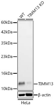 | Western blot analysis of lysates from wild type (WT) and TIMM13 knockdown (KD) HeLa cells using [KD Validated] TIMM13 Rabbit mAb (A25135) at 1:1000 dilution. Secondary antibody: HRP-conjugated Goat anti-Rabbit IgG (H+L) (AS014) at 1:10000 dilution.Lysates/proteins: 25 μg per lane. Blocking buffer: 3% nonfat dry milk in TBST. Detection: ECL Basic Kit (RM00020). Exposure time: 90s. |
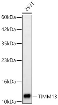 | Western blot analysis of lysates from 293T cells using [KD Validated] TIMM13 Rabbit mAb (A25135) at 1:1000 dilution. Secondary antibody: HRP-conjugated Goat anti-Rabbit IgG (H+L) (AS014) at 1:10000 dilution. Lysates/proteins: 25 μg per lane. Blocking buffer: 3% nonfat dry milk in TBST. Detection: ECL Basic Kit (RM00020). Exposure time: 90s. |
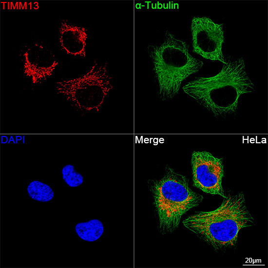 | Confocal imaging of HeLa cells using [KD Validated] TIMM13 Rabbit mAb (A25135,dilution 1:200) followed by a further incubation with Cy3 Goat Anti-Rabbit IgG (H+L) (AS007,dilution 1:500)(Red).The cells were counterstained with α-Tubulin Mouse mAb (AC012, dilution 1:400) followed by incubation with ABflo® 488-conjugated Goat Anti-Mouse IgG (H+L) Ab (AS076, dilution 1:500) (Green).DAPI was used for nuclear staining (Blue). Objective: 100x. |
You may also be interested in:

