Your shopping cart is empty!
| Reactivity: | Human |
| Applications: | WB, ELISA |
| Host Species: | Rabbit |
| Isotype: | IgG |
| Clonality: | Polyclonal antibody |
| Gene Name: | ubiquitin like with PHD and ring finger domains 1 |
| Gene Symbol: | UHRF1 |
| Synonyms: | Np95; hNP95; ICBP90; RNF106; TDRD22; hUHRF1; huNp95 |
| Gene ID: | 29128 |
| UniProt ID: | Q96T88 |
| Immunogen: | Recombinant fusion protein containing a sequence corresponding to amino acids 672-793 of human UHRF1 (NP_037414.3). |
| Dilution: | WB 1:500-1:1000; IHC 1:50-1:200 |
| Purification Method: | Affinity purification |
| Concentration: | 0.55 mg/mL |
| Buffer: | PBS with 0.05% proclin300, 50% glycerol, pH7.3. |
| Storage: | Store at -20°C. Avoid freeze / thaw cycles. |
| Documents: | Manual-UHRF1 polyclonal antibody |
Background
This gene encodes a member of a subfamily of RING-finger type E3 ubiquitin ligases. The protein binds to specific DNA sequences, and recruits a histone deacetylase to regulate gene expression. Its expression peaks at late G1 phase and continues during G2 and M phases of the cell cycle. It plays a major role in the G1/S transition by regulating topoisomerase IIalpha and retinoblastoma gene expression, and functions in the p53-dependent DNA damage checkpoint. It is regarded as a hub protein for the integration of epigenetic information. This gene is up-regulated in various cancers, and it is therefore considered to be a therapeutic target. Multiple transcript variants encoding different isoforms have been found for this gene. A related pseudogene exists on chromosome 12.
Images
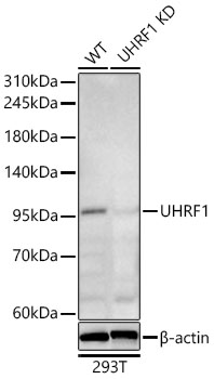 | Western blot analysis of lysates from wild type (WT) and UHRF1 knockdown (KD) 293T cells using [KD Validated] UHRF1 Rabbit pAb (A25717) at 1:1000 dilution incubated overnight at 4°C. Secondary antibody: HRP-conjugated Goat anti-Rabbit IgG (H+L) (AS014) at 1:10000 dilution. Lysates/proteins: 25 μg per lane. Blocking buffer: 3% nonfat dry milk in TBST. Detection: ECL Basic Kit (RM00020) Exposure time: 180 s. |
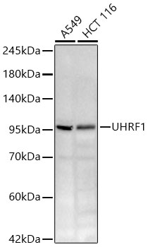 | Western blot analysis of various lysates using [KD Validated] UHRF1 Rabbit pAb (A25717) at 1:1000 dilution incubated overnight at 4°C. Secondary antibody: HRP-conjugated Goat anti-Rabbit IgG (H+L) (AS014) at 1:10000 dilution. Lysates/proteins: 25 μg per lane. Blocking buffer: 3% nonfat dry milk in TBST. Detection: ECL Basic Kit (RM00020) Exposure time: 180 s. |
You may also be interested in:

