Your shopping cart is empty!
")
| Reactivity: | Human, Mouse, Rat |
| Applications: | WB, IHC, IF/IC, IP, ELISA |
| Host Species: | Rabbit |
| Isotype: | IgG |
| Clonality: | Monoclonal antibody |
| Gene Name: | upstream transcription factor 1 |
| Gene Symbol: | USF1 |
| Synonyms: | UEF; FCHL; MLTF; FCHL1; MLTFI; HYPLIP1; bHLHb11; [KD Validated] USF1 |
| Gene ID: | 7391 |
| UniProt ID: | P22415 |
| Clone ID: | 6T8L3 |
| Immunogen: | Recombinant fusion protein containing a sequence corresponding to amino acids 1-310 of human USF1. (NP_009053.1). |
| Dilution: | WB 1:2000-1:10000; IHC 1:500-1:1000; IF/IC 1:50-1:200 |
| Purification Method: | Affinity purification |
| Concentration: | 0.58 mg/mL |
| Buffer: | PBS with 0.05% proclin300, 0.05% BSA, 50% glycerol, pH7.3. |
| Storage: | Store at -20°C. Avoid freeze / thaw cycles. |
| Documents: | Manual-USF1 monoclonal antibody |
Background
This gene encodes a member of the basic helix-loop-helix leucine zipper family, and can function as a cellular transcription factor. The encoded protein can activate transcription through pyrimidine-rich initiator (Inr) elements and E-box motifs. This gene has been linked to familial combined hyperlipidemia (FCHL). Alternative splicing of this gene results in multiple transcript variants. A related pseudogene has been defined on chromosome 21.
Images
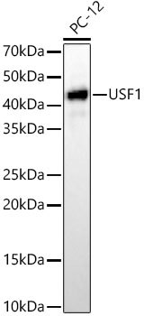 | Western blot analysis of lysates from PC-12 cells, using [KD Validated] USF1 Rabbit mAb (A23474) at 1:10000 dilution. Secondary antibody: HRP-conjugated Goat anti-Rabbit IgG (H+L) (AS014) at 1:10000 dilution. Lysates/proteins: 25μg per lane. Blocking buffer: 3% nonfat dry milk in TBST. Detection: ECL Basic Kit (RM00020). Exposure time: 60s. |
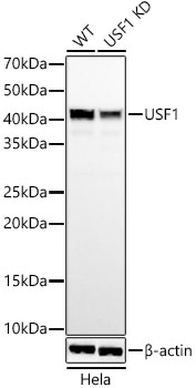 | Western blot analysis of lysates from wild type(WT) and USF1 knockdown (KD) Hela cells, using [KD Validated] USF1 Rabbit mAb (A23474) at 1:10000 dilution. Secondary antibody: HRP-conjugated Goat anti-Rabbit IgG (H+L) (AS014) at 1:10000 dilution. Lysates/proteins: 25μg per lane. Blocking buffer: 3% nonfat dry milk in TBST. Detection: ECL Basic Kit (RM00020). Exposure time: 60s. |
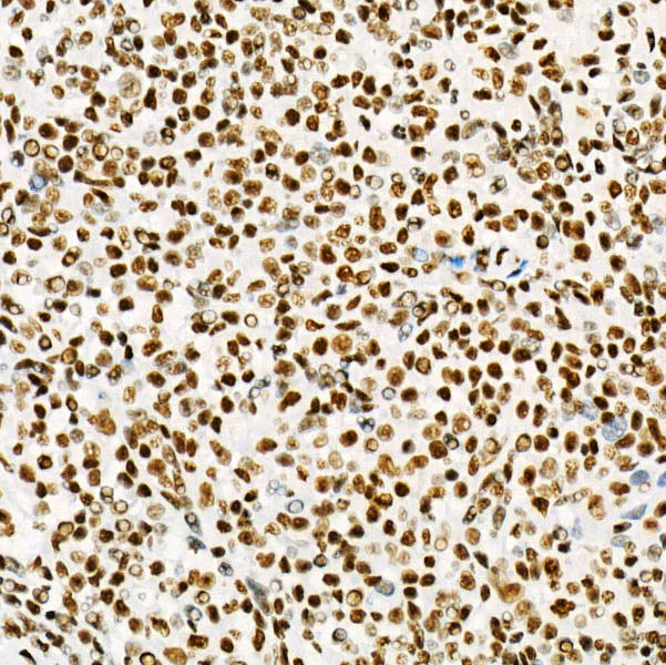 | Immunohistochemistry analysis of paraffin-embedded Human spleen using USF1 Rabbit mAb (A23474) at dilution of 1:900 (40x lens). High pressure antigen retrieval performed with 0.01M Citrate Bufferr (pH 6.0) prior to IHC staining. |
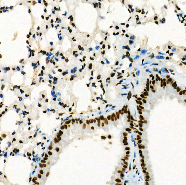 | Immunohistochemistry analysis of paraffin-embedded Mouse lung using USF1 Rabbit mAb (A23474) at dilution of 1:900 (40x lens). High pressure antigen retrieval performed with 0.01M Citrate Bufferr (pH 6.0) prior to IHC staining. |
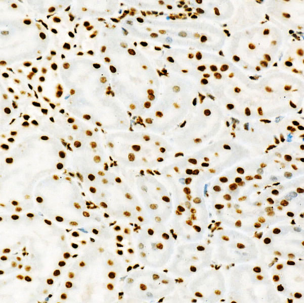 | Immunohistochemistry analysis of paraffin-embedded Rat kidney using USF1 Rabbit mAb (A23474) at dilution of 1:900 (40x lens). High pressure antigen retrieval performed with 0.01M Citrate Bufferr (pH 6.0) prior to IHC staining. |
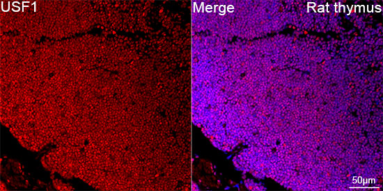 | Confocal imaging of paraffin-embedded Rat thymus using [KD Validated] USF1 Rabbit mAb (A23474,dilution 1:200) followed by a further incubation with Cy3 Goat Anti-Rabbit IgG (H+L) (AS007,dilution 1:500)(Red).DAPI was used for nuclear staining (Blue). Objective: 40x. Perform high pressure antigen retrieval with 0.01 M citRate buffer (pH 6.0) prior to IF staining. |
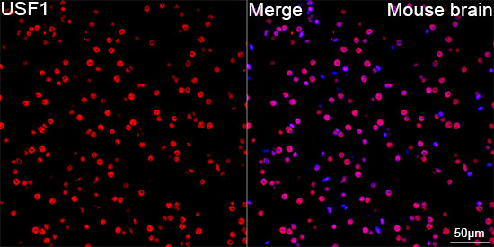 | Confocal imaging of paraffin-embedded Mouse brain using [KD Validated] USF1 Rabbit mAb (A23474,dilution 1:200) followed by a further incubation with Cy3 Goat Anti-Rabbit IgG (H+L) (AS007,dilution 1:500)(Red).DAPI was used for nuclear staining (Blue). Objective: 40x. |
You may also be interested in:

