Your shopping cart is empty!
KD-Validated YTHDC1 Rabbit mAb (20 μl)
| Reactivity: | Human |
| Applications: | WB, IHC-P, IP, ELISA |
| Host Species: | Rabbit |
| Isotype: | IgG |
| Clonality: | Monoclonal antibody |
| Gene Name: | YTH N6-methyladenosine RNA binding protein C1 |
| Gene Symbol: | YTHDC1 |
| Synonyms: | YT521; YT521-B; [KD Validated] YTHDC1 |
| Gene ID: | 91746 |
| UniProt ID: | Q96MU7 |
| Clone ID: | 9M7W2 |
| Immunogen: | Recombinant fusion protein containing a sequence corresponding to amino acids 1-71 of human YTHDC1 (NP_001026902.1). |
| Dilution: | WB 1:500-1:1000 |
| Purification Method: | Affinity purification |
| Concentration: | 1.34 mg/ml |
| Buffer: | PBS with 0.05% proclin300, 0.05% BSA, 50% glycerol, pH7.3. |
| Storage: | Store at -20°C. Avoid freeze / thaw cycles. |
| Documents: | Manual-YTHDC1 monoclonal antibody |
Background
Enables N6-methyladenosine-containing RNA binding activity. Involved in mRNA export from nucleus; mRNA splice site selection; and regulation of gene expression. Located in nuclear speck and plasma membrane.
Images
 | Western blot analysis of various lysates using [KD Validated] YTHDC1 Rabbit mAb (A22992) at 1:2000 dilution incubated overnight at 4℃. Secondary antibody: HRP-conjugated Goat anti-Rabbit IgG (H+L) (AS014) at 1:10000 dilution. Lysates/proteins: 25μg per lane. Blocking buffer: 3% nonfat dry milk in TBST. Detection: ECL Basic Kit (RM00020). Exposure time: 30s. |
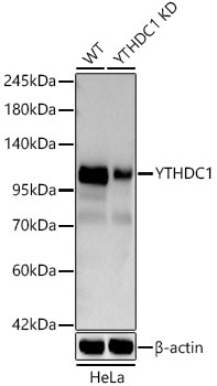 | Western blot analysis of lysates from wild type (WT) and YTHDC1 knockdown (KD) HeLa cells using [KD Validated] YTHDC1 Rabbit mAb (A22992) at 1:3000 dilution incubated overnight at 4℃. Secondary antibody: HRP-conjugated Goat anti-Rabbit IgG (H+L) (AS014) at 1:10000 dilution. Lysates/proteins: 25ug per lane. Blocking buffer: 3% nonfat dry milk in TBST. Detection: ECL Basic Kit (RM00020). Exposure time: 30s. |
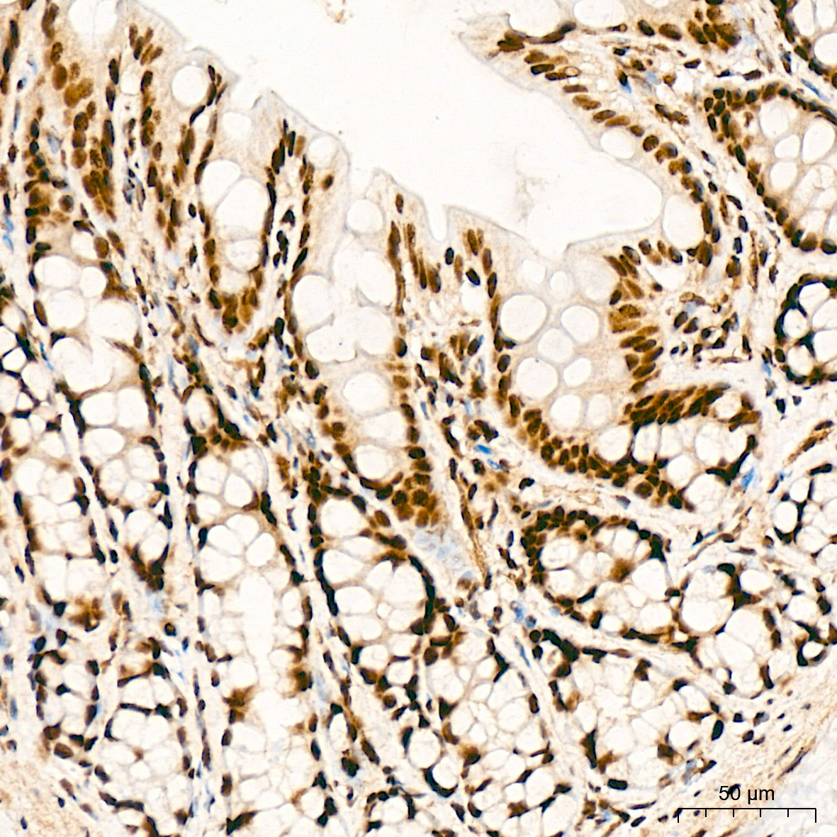
 | Immunohistochemistry analysis of paraffin-embedded Mouse colon tissue using [KD Validated] YTHDC1 Rabbit mAb (A22992) at a dilution of 1:1000 (40x lens). High pressure antigen retrieval performed with 0.01M Citrate Buffer (pH 6.0) prior to IHC staining. |
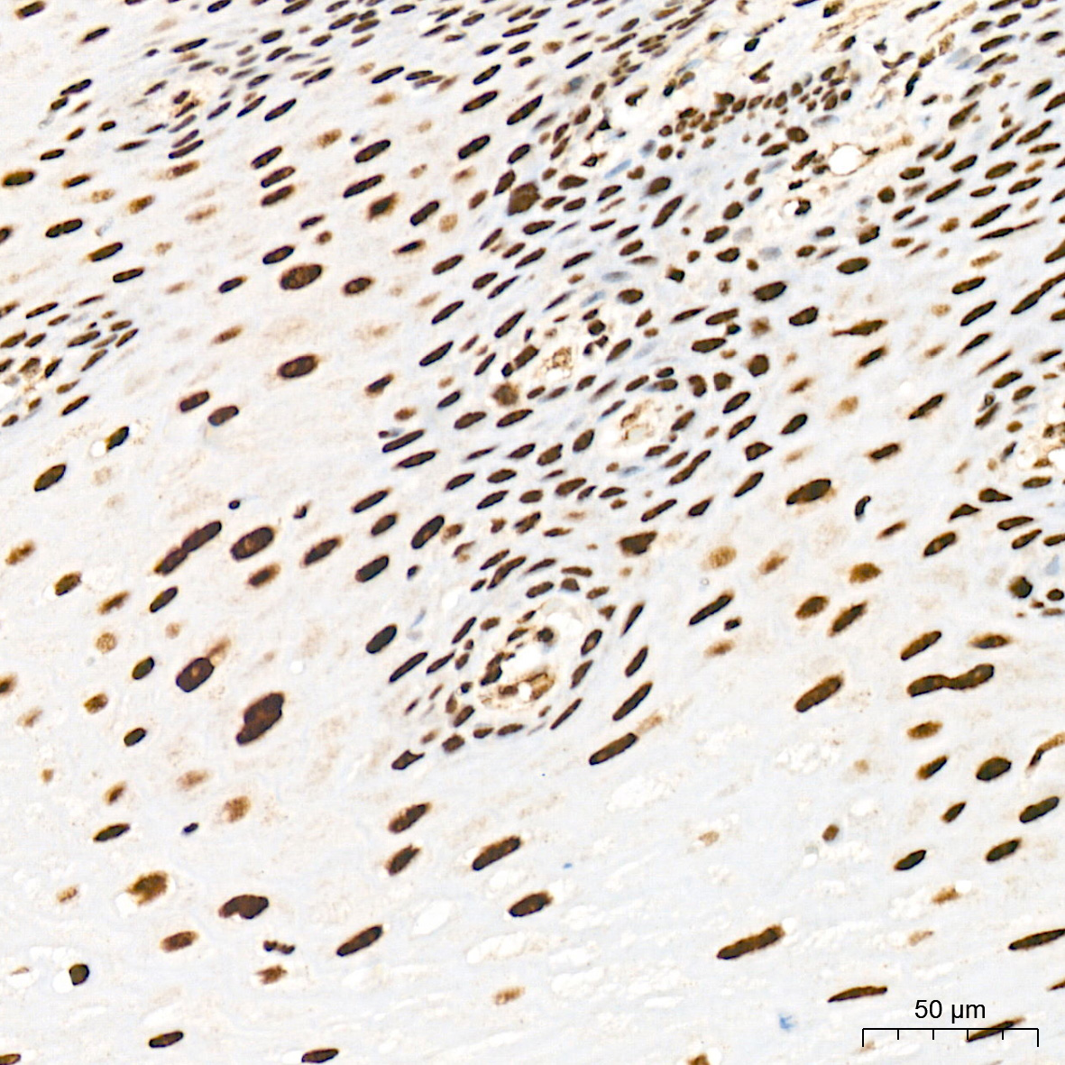
 | Immunohistochemistry analysis of paraffin-embedded Human thyroid cancer tissue using [KD Validated] YTHDC1 Rabbit mAb (A22992) at a dilution of 1:1000 (40x lens). High pressure antigen retrieval performed with 0.01M Citrate Buffer (pH 6.0) prior to IHC staining. |
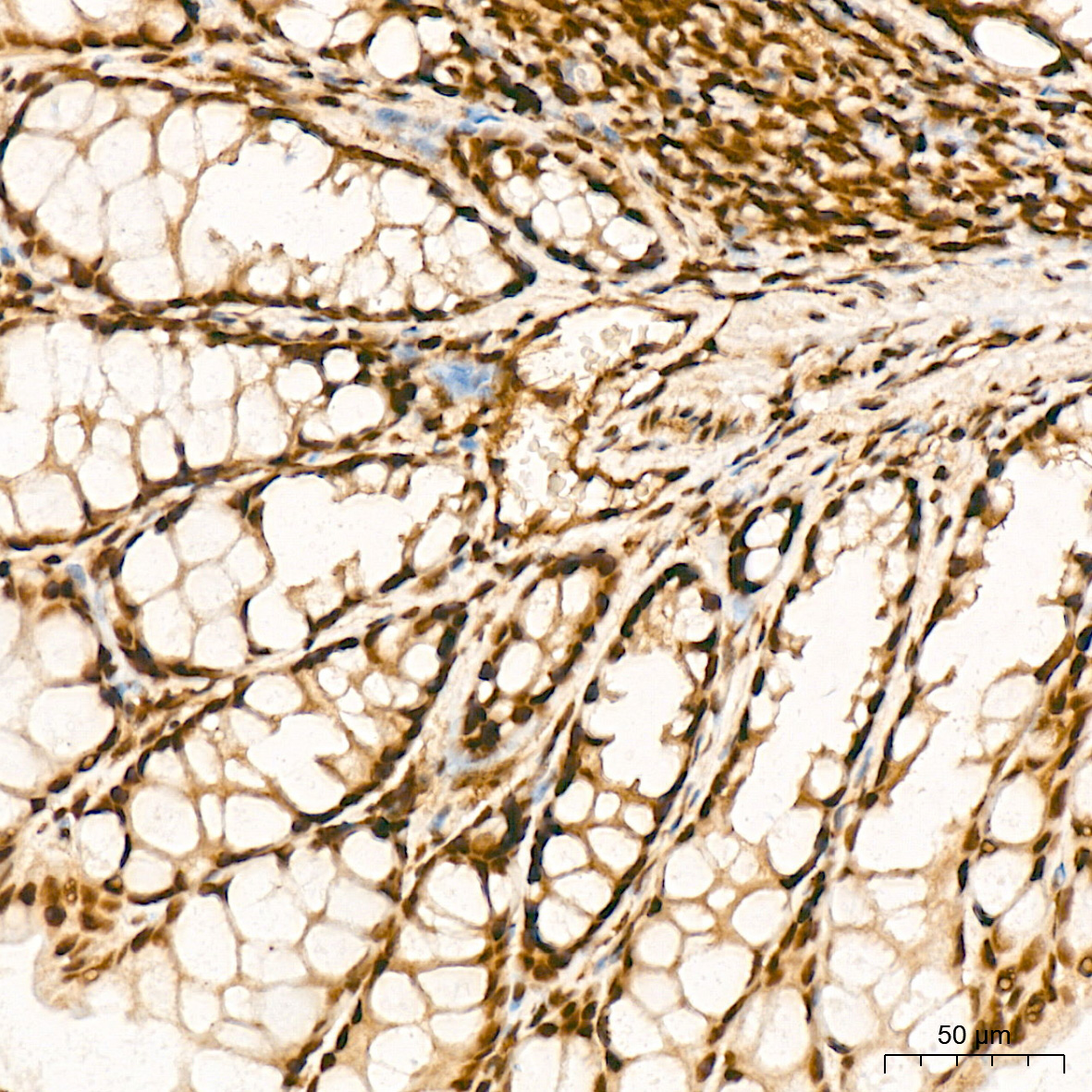
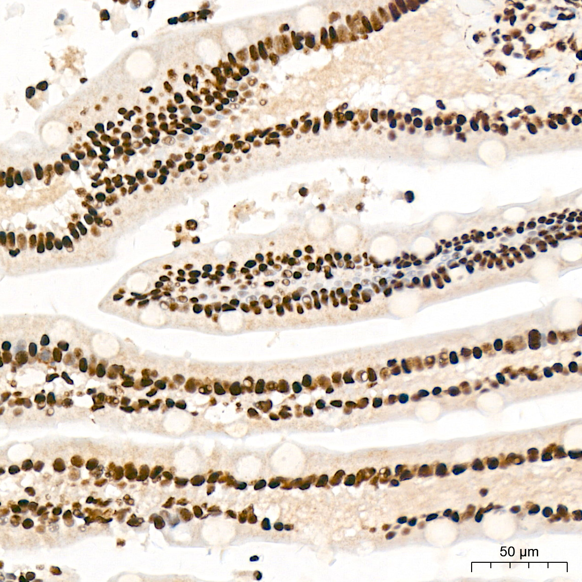
 | Immunohistochemistry analysis of paraffin-embedded Rat colon tissue using [KD Validated] YTHDC1 Rabbit mAb (A22992) at a dilution of 1:1000 (40x lens). High pressure antigen retrieval performed with 0.01M Citrate Buffer (pH 6.0) prior to IHC staining. |
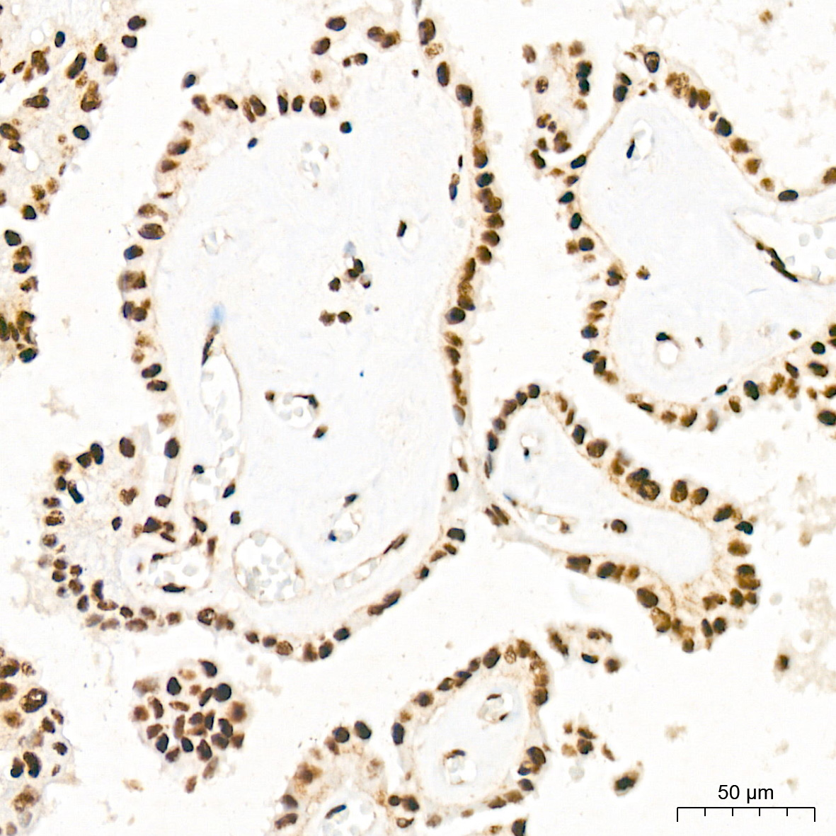
 | Immunohistochemistry analysis of paraffin-embedded Human small intestine tissue using [KD Validated] YTHDC1 Rabbit mAb (A22992) at a dilution of 1:1000 (40x lens). High pressure antigen retrieval performed with 0.01M Citrate Buffer (pH 6.0) prior to IHC staining. |
 | Immunohistochemistry analysis of paraffin-embedded Human esophagus tissue using [KD Validated] YTHDC1 Rabbit mAb (A22992) at a dilution of 1:1000 (40x lens). High pressure antigen retrieval performed with 0.01M Citrate Buffer (pH 6.0) prior to IHC staining. |
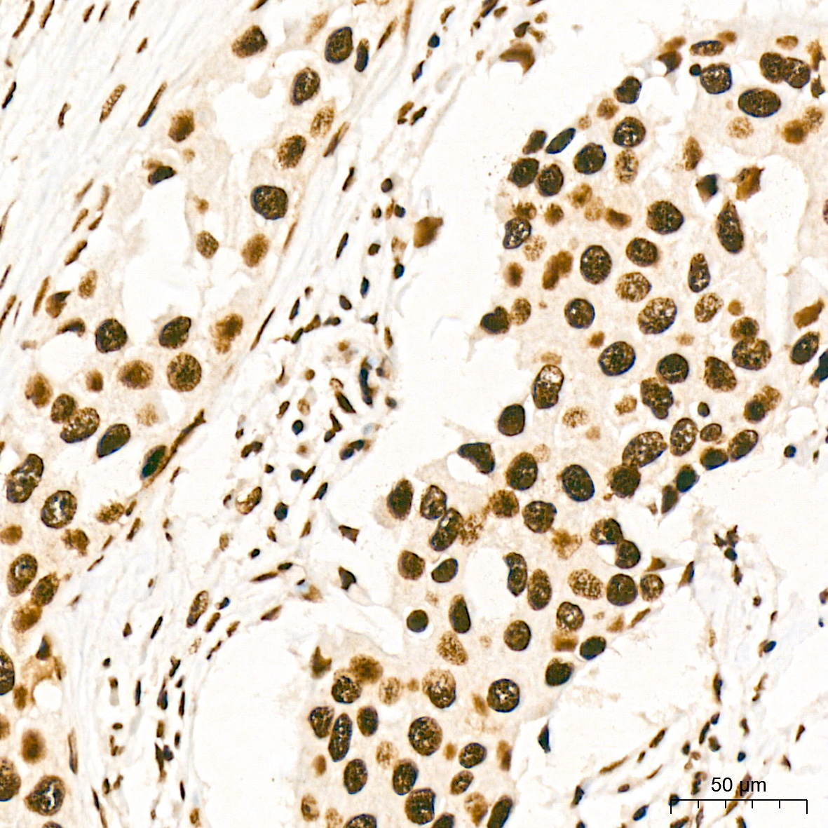 | Immunohistochemistry analysis of paraffin-embedded Human breast cancer tissue using [KD Validated] YTHDC1 Rabbit mAb (A22992) at a dilution of 1:1000 (40x lens). High pressure antigen retrieval performed with 0.01M Citrate Buffer (pH 6.0) prior to IHC staining. |
You may also be interested in:

