Your shopping cart is empty!
KO-Validated ATG16L1 Rabbit mAb (20 μl)
| Reactivity: | Human |
| Applications: | WB, ELISA |
| Host Species: | Rabbit |
| Isotype: | IgG |
| Clonality: | Monoclonal antibody |
| Gene Name: | autophagy related 16 like 1 |
| Gene Symbol: | ATG16L1 |
| Synonyms: | IBD10; WDR30; APG16L; ATG16A; ATG16L; ATG16L1 |
| Gene ID: | 55054 |
| UniProt ID: | Q676U5 |
| Clone ID: | 5N2N9 |
| Immunogen: | Recombinant fusion protein containing a sequence corresponding to amino acids 6-105 of human ATG16L1 (Q676U5). |
| Dilution: | WB 1:500-1:2000; IF/IC 1:50-1:100 |
| Purification Method: | Affinity purification |
| Concentration: | 1 mg/ml |
| Buffer: | PBS with 0.02% sodium azide, 0.05% BSA, 50% glycerol, pH7.3. |
| Storage: | Store at -20°C. Avoid freeze / thaw cycles. |
| Documents: | Manual-ATG16L1 monoclonal antibody |
Background
The protein encoded by this gene is part of a large protein complex that is necessary for autophagy, the major process by which intracellular components are targeted to lysosomes for degradation. Defects in this gene are a cause of susceptibility to inflammatory bowel disease type 10 (IBD10). Several transcript variants encoding different isoforms have been found for this gene.
Images
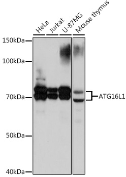 | Western blot analysis of various lysates using [KO Validated] ATG16L1 Rabbit mAb (A3637) at 1:1000 dilution. Secondary antibody: HRP-conjugated Goat anti-Rabbit IgG (H+L) (AS014) at 1:10000 dilution. Lysates/proteins: 25μg per lane. Blocking buffer: 3% nonfat dry milk in TBST. Detection: ECL Basic Kit (RM00020). Exposure time: 30s. |
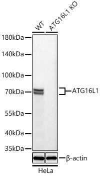 | Western blot analysis of lysates from wild type (WT) and ATG16L1 knockout (KO) HeLa cells using [KO Validated] ATG16L1 Rabbit mAb (A3637) at 1:1000 dilution incubated overnight at 4℃. Secondary antibody: HRP-conjugated Goat anti-Rabbit IgG (H+L) (AS014) at 1:10000 dilution. Lysates/proteins: 25 μg per lane. Blocking buffer: 3% nonfat dry milk in TBST. Detection: ECL Basic Kit (RM00020). Exposure time: 10s. |
You may also be interested in:

