Your shopping cart is empty!
KO-Validated CDKN1A/p21 Rabbit PolymAb® (20 μl)
| Reactivity: | Human, Mouse |
| Applications: | WB, IF/ICC, ELISA |
| Host Species: | Rabbit |
| Isotype: | IgG |
| Clonality: | Monoclonal antibody |
| Gene Name: | cyclin dependent kinase inhibitor 1A |
| Gene Symbol: | CDKN1A |
| Synonyms: | P21; CIP1; SDI1; WAF1; CAP20; CDKN1; MDA-6; p21CIP1 |
| Gene ID: | 1026/12575 |
| UniProt ID: | P38936/P39689 |
| Clone ID: | 4F3R2 |
| Immunogen: | Recombinant protein of human/mouse CDKN1A/p21. |
| Purification Method: | Affinity purification |
| Concentration: | 0.97 mg/ml |
| Buffer: | PBS with 0.05% proclin300, 0.05% BSA, 50% glycerol, pH7.3. |
| Storage: | Store at -20°C. Avoid freeze / thaw cycles. |
| Documents: | Manual-CDKN1A/p21 monoclonal antibody |
Background
This gene encodes a potent cyclin-dependent kinase inhibitor. The encoded protein binds to and inhibits the activity of cyclin-cyclin-dependent kinase2 or -cyclin-dependent kinase4 complexes, and thus functions as a regulator of cell cycle progression at G1. The expression of this gene is tightly controlled by the tumor suppressor protein p53, through which this protein mediates the p53-dependent cell cycle G1 phase arrest in response to a variety of stress stimuli. This protein can interact with proliferating cell nuclear antigen, a DNA polymerase accessory factor, and plays a regulatory role in S phase DNA replication and DNA damage repair. This protein was reported to be specifically cleaved by CASP3-like caspases, which thus leads to a dramatic activation of cyclin-dependent kinase2, and may be instrumental in the execution of apoptosis following caspase activation. Mice that lack this gene have the ability to regenerate damaged or missing tissue. Multiple alternatively spliced variants have been found for this gene.
Images
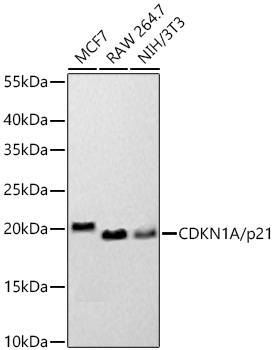 | Western blot analysis of various lysates using [KO Validated] CDKN1A/p21 Rabbit PolymAb® (A22460PM) at 1:20000 dilution incubated overnight at 4℃. Secondary antibody: HRP-conjugated Goat anti-Rabbit IgG (H+L) (AS014) at 1:10000 dilution. Lysates/proteins: 25 μg per lane. Blocking buffer: 3% nonfat dry milk in TBST. Detection: ECL Basic Kit (RM00020). Exposure time: 45s. |
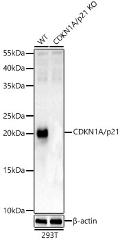 | Western blot analysis of lysates from wild type(WT) and CDKN1A/p21 knockout (KO) 293T cells, using [KO Validated] CDKN1A/p21 Rabbit PolymAb® (A22460PM) at 1:500 dilution. Secondary antibody: HRP-conjugated Goat anti-Rabbit IgG (H+L) (AS014) at 1:10000 dilution. Lysates/proteins: 25μg per lane. Blocking buffer: 3% nonfat dry milk in TBST. Detection: ECL Basic Kit (RM00020). Exposure time: 180s. |
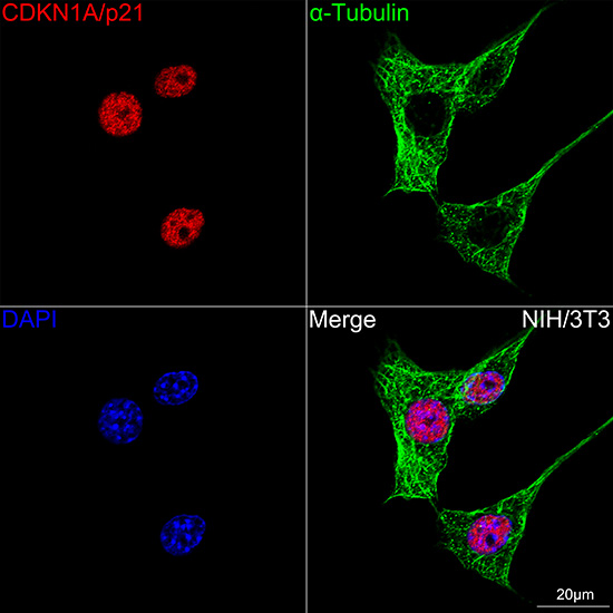 | Confocal imaging of NIH/3T3 cells using [KO Validated] CDKN1A/p21 Rabbit PolymAb® (A22460PM,dilution 1:1000)(Red) followed by a further incubation with Cy3 Goat Anti-Rabbit IgG (H+L) (AS007, dilution 1:500) (Red). The cells were counterstained with α-tubulin Mouse mAb (AC012, dilution 1:400) followed by incubation with ABflo® 488-conjugated Goat Anti-Mouse IgG (H+L) Ab (AS076, dilution 1:500) (Green). DAPI was used for nuclear staining (Blue). Objective: 100x. |
You may also be interested in:

