Your shopping cart is empty!
KO-Validated PHD2/EGLN1 Rabbit pAb (20 μl)
| Reactivity: | Human |
| Applications: | WB, IHC-P, IF/ICC, ELISA |
| Host Species: | Rabbit |
| Isotype: | IgG |
| Clonality: | Polyclonal antibody |
| Gene Name: | egl-9 family hypoxia inducible factor 1 |
| Gene Symbol: | EGLN1 |
| Synonyms: | HPH2; PHD2; SM20; ECYT3; HALAH; HPH-2; HIFPH2; ZMYND6; C1orf12; HIF-PH2; N1 |
| Gene ID: | 54583 |
| UniProt ID: | Q9GZT9 |
| Immunogen: | A synthetic peptide corresponding to a sequence within amino acids 1-100 of human PHD2/EGLN1 (NP_071334.1). |
| Dilution: | WB 1:500-1:2000 |
| Purification Method: | Affinity purification |
| Concentration: | 0.69 mg/ml |
| Buffer: | PBS with 0.05% proclin300, 50% glycerol, pH7.3. |
| Storage: | Store at -20°C. Avoid freeze / thaw cycles. |
| Documents: | Manual-EGLN1 polyclonal antibody |
Background
The protein encoded by this gene catalyzes the post-translational formation of 4-hydroxyproline in hypoxia-inducible factor (HIF) alpha proteins. HIF is a transcriptional complex that plays a central role in mammalian oxygen homeostasis. This protein functions as a cellular oxygen sensor, and under normal oxygen concentration, modification by prolyl hydroxylation is a key regulatory event that targets HIF subunits for proteasomal destruction via the von Hippel-Lindau ubiquitylation complex. Mutations in this gene are associated with erythrocytosis familial type 3 (ECYT3).
Images
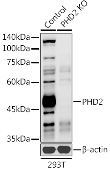 | Western blot analysis of lysates from wild type (WT) and PHD2/EGLN1 knockout (KO) 293T cells, using [KO Validated] PHD2/EGLN1 Rabbit pAb (A14557) at 1:1000 dilution. Secondary antibody: HRP-conjugated Goat anti-Rabbit IgG (H+L) (AS014) at 1:10000 dilution. Lysates/proteins: 25μg per lane. Blocking buffer: 3% nonfat dry milk in TBST. Detection: ECL Basic Kit (RM00020). Exposure time: 90s. |
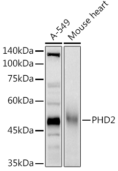 | Western blot analysis of various lysates using [KO Validated] PHD2/EGLN1 Rabbit pAb (A14557) at 1:1000 dilution. Secondary antibody: HRP-conjugated Goat anti-Rabbit IgG (H+L) (AS014) at 1:10000 dilution. Lysates/proteins: 25μg per lane. Blocking buffer: 3% nonfat dry milk in TBST. Detection: ECL Basic Kit (RM00020). Exposure time: 90s. |
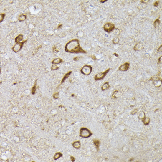 | Immunohistochemistry analysis of paraffin-embedded Mouse brain using [KO Validated] PHD2/EGLN1 Rabbit pAb (A14557) at dilution of 1:100 (40x lens). High pressure antigen retrieval performed with 0.01M Citrate Bufferr (pH 6.0) prior to IHC staining. |
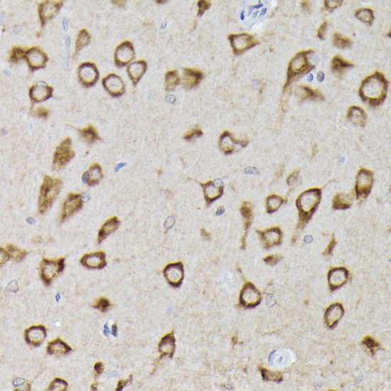 | Immunohistochemistry analysis of paraffin-embedded Rat brain using [KO Validated] PHD2/EGLN1 Rabbit pAb (A14557) at dilution of 1:100 (40x lens). High pressure antigen retrieval performed with 0.01M Citrate Bufferr (pH 6.0) prior to IHC staining. |
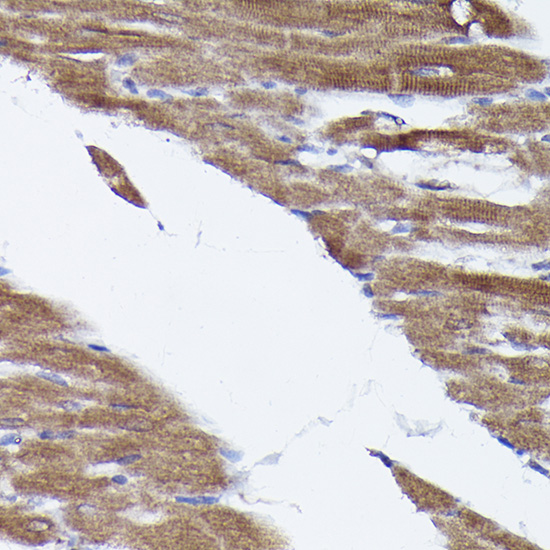 | Immunohistochemistry analysis of paraffin-embedded Rat heart using [KO Validated] PHD2/EGLN1 Rabbit pAb (A14557) at dilution of 1:100 (40x lens). High pressure antigen retrieval performed with 0.01M Citrate Bufferr (pH 6.0) prior to IHC staining. |
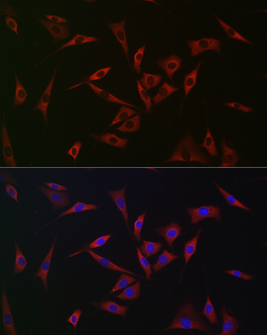 | Immunofluorescence analysis of NIH/3T3 cells using [KO Validated] PHD2/EGLN1 Rabbit pAb (A14557) at dilution of 1:100 (40x lens). Secondary antibody: Cy3-conjugated Goat anti-Rabbit IgG (H+L) (AS007) at 1:500 dilution. Blue: DAPI for nuclear staining. |
You may also be interested in:

