Your shopping cart is empty!
KO-Validated TTC11/FIS1 Rabbit mAb (20 μl)
| Reactivity: | Human |
| Applications: | WB, IHC-P, IF/ICC, IP, ELISA |
| Host Species: | Rabbit |
| Isotype: | IgG |
| Clonality: | Monoclonal antibody |
| Gene Name: | fission, mitochondrial 1 |
| Gene Symbol: | FIS1 |
| Synonyms: | TTC11; CGI-135; S1 |
| Gene ID: | 51024 |
| UniProt ID: | Q9Y3D6 |
| Clone ID: | 6S1G4 |
| Immunogen: | Recombinant fusion protein containing a sequence corresponding to amino acids 1-122 of human TTC11/FIS1 (NP_057152.2). |
| Dilution: | WB 1:2000-1:20000 |
| Purification Method: | Affinity purification |
| Concentration: | 1 mg/ml |
| Buffer: | PBS with 0.05% proclin300, 0.05% BSA, 50% glycerol, pH7.3. |
| Storage: | Store at -20°C. Avoid freeze / thaw cycles. |
| Documents: | Manual-FIS1 monoclonal antibody |
Background
Enables identical protein binding activity. Involved in several processes, including calcium-mediated signaling using intracellular calcium source; cellular calcium ion homeostasis; and mitochondrion organization. Acts upstream of or within mitochondrion morphogenesis. Located in mitochondrion and peroxisome. Is integral component of mitochondrial outer membrane and integral component of peroxisomal membrane. Part of protein-containing complex. Biomarker of Alzheimer's disease.
Images
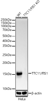 | Western blot analysis of lysates from wild type (WT) and TTC11/FIS1 knockout (KO) HeLa(KO) cells, using [KO Validated] TTC11/FIS1 Rabbit mAb (A19666) at 1:1000 dilution incubated overnight at 4℃. Secondary antibody: HRP-conjugated Goat anti-Rabbit IgG (H+L) (AS014) at 1:10000 dilution. Lysates/proteins: 25μg per lane. Blocking buffer: 3% nonfat dry milk in TBST. Detection: ECL Basic Kit (RM00020). Exposure time: 10s. |
 | Western blot analysis of various lysates, using [KO Validated] TTC11/FIS1 Rabbit mAb (A19666) at 1:1000 dilution incubated overnight at 4℃. Secondary antibody: HRP-conjugated Goat anti-Rabbit IgG (H+L) (AS014) at 1:10000 dilution. Lysates/proteins: 25μg per lane. Blocking buffer: 3% nonfat dry milk in TBST. Detection: ECL Basic Kit (RM00020). Exposure time: 10s. |
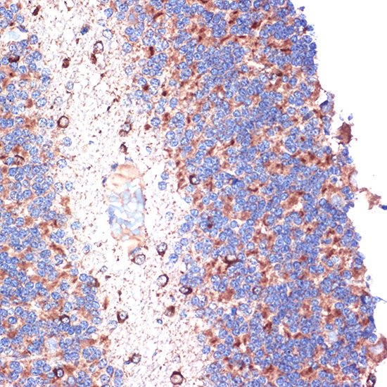 | Immunohistochemistry analysis of paraffin-embedded Rat brain tissue using [KO Validated] TTC11/FIS1 Rabbit mAb (A19666) at a dilution of 1:100 (40x lens). Microwave antigen retrieval performed with 0.01M Tris-EDTA Buffer (pH 9.0) prior to IHC staining. |
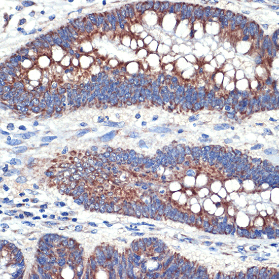 | Immunohistochemistry analysis of paraffin-embedded Human colon carcinoma tissue using [KO Validated] TTC11/FIS1 Rabbit mAb (A19666) at a dilution of 1:100 (40x lens). Microwave antigen retrieval performed with 0.01M Tris-EDTA Buffer (pH 9.0) prior to IHC staining. |
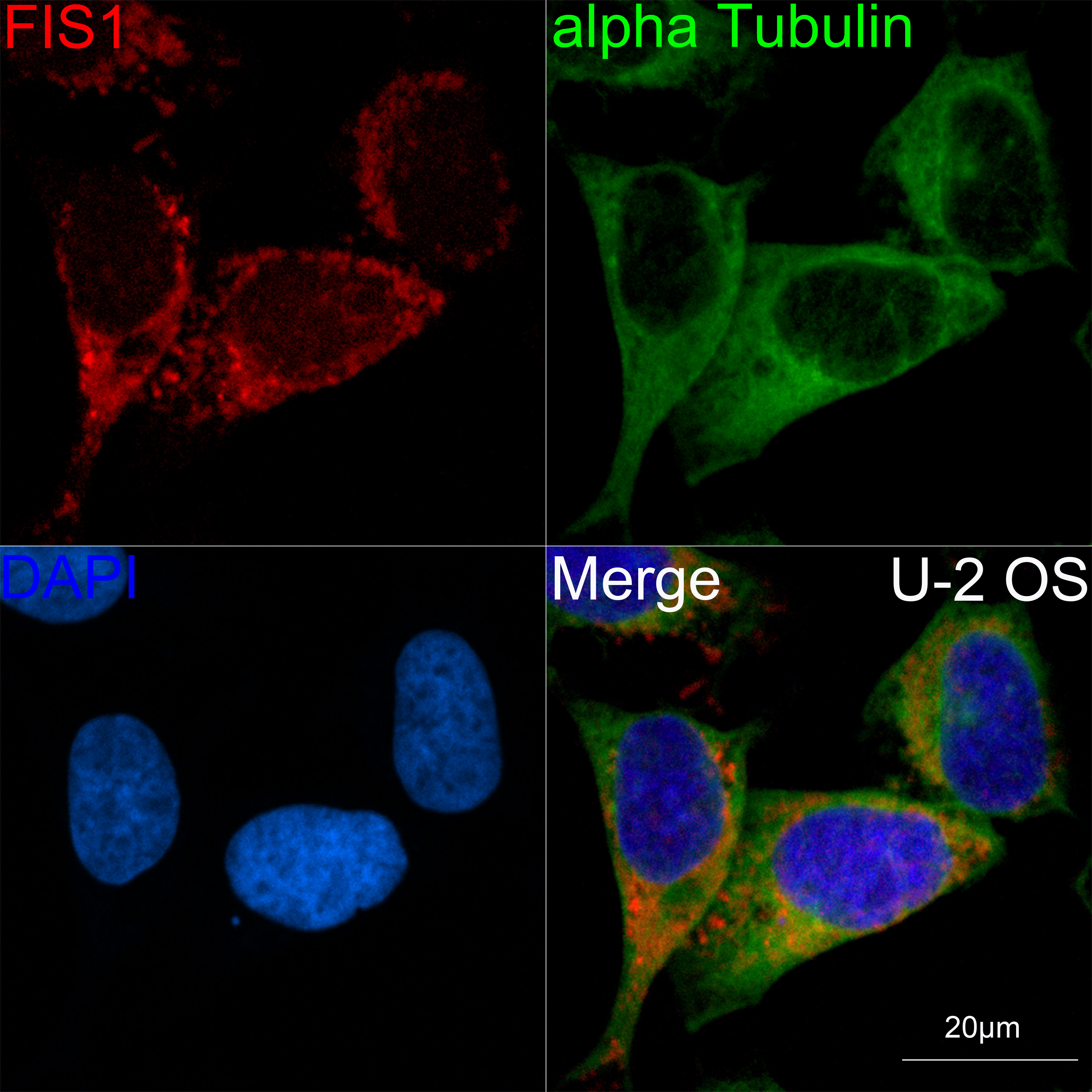 | Confocal imaging of U-2 OS cells using [KO Validated] TTC11/FIS1 Rabbit mAb (A19666,dilution 1:100)(Red). The cells were counterstained with α-Tubulin Mouse mAb (AC012,dilution 1:400) (Green). DAPI was used for nuclear staining (blue). Objective: 60x. |
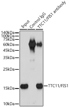 | Immunoprecipitation of TTC11/FIS1 from 300 µg extracts of HeLa cells was performed using 3 µg of TTC11/FIS1 antibody (A19666). Rabbit IgG isotype control (AC042) was used to precipitate the Control IgG sample. IP samples were eluted with 1X reducing Laemmli Buffer. The Input lane represents 10% of the total input. Western blot analysis of immunoprecipitates was conducted using TTC11/FIS1 (A19666) at a dilution of 1:2000. |
You may also be interested in:

