Your shopping cart is empty!
")
KO-Validated Fascin/FSCN1 Rabbit pAb (20 μl)
| Reactivity: | Human, Mouse, Rat |
| Applications: | WB, IHC, IF/IC, ELISA |
| Host Species: | Rabbit |
| Isotype: | IgG |
| Clonality: | Polyclonal antibody |
| Gene Name: | fascin actin-bundling protein 1 |
| Gene Symbol: | FSCN1 |
| Synonyms: | HSN; SNL; p55; FAN1; N1 |
| Gene ID: | 6624 |
| UniProt ID: | Q16658 |
| Immunogen: | Recombinant fusion protein containing a sequence corresponding to amino acids 394-493 of human Fascin/Fascin/FSCN1 (NP_003079.1). |
| Dilution: | WB 1:500-1:2000; IHC 1:50-1:200; IF/IC 1:50-1:200 |
| Purification Method: | Affinity purification |
| Concentration: | 0.69 mg/ml |
| Buffer: | PBS with 0.02% sodium azide, 50% glycerol ,pH7.3. |
| Storage: | Store at -20°C. Avoid freeze / thaw cycles. |
| Documents: | Manual-FSCN1 polyclonal antibody |
Background
This gene encodes a member of the fascin family of actin-binding proteins. Fascin proteins organize F-actin into parallel bundles, and are required for the formation of actin-based cellular protrusions. The encoded protein plays a critical role in cell migration, motility, adhesion and cellular interactions. Expression of this gene is known to be regulated by several microRNAs, and overexpression of this gene may play a role in the metastasis of multiple types of cancer by increasing cell motility. Expression of this gene is also a marker for Reed-Sternberg cells in Hodgkin's lymphoma. A pseudogene of this gene is located on the long arm of chromosome 15.
Images
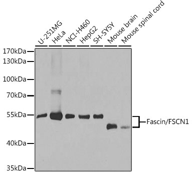 | Western blot analysis of various lysates using [KO Validated] Fascin/FSCN1 Rabbit pAb (A1904) at 1:1000 dilution. Secondary antibody: HRP-conjugated Goat anti-Rabbit IgG (H+L) (AS014) at 1:10000 dilution. Lysates/proteins: 25μg per lane. Blocking buffer: 3% nonfat dry milk in TBST. Detection: ECL Basic Kit (RM00020). Exposure time: 30s. |
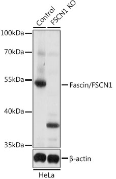 | Western blot analysis of lysates from wild type (WT) and Fascin/Fascin/FSCN1 knockout (KO) HeLa cells, using [KO Validated] Fascin/FSCN1 Rabbit pAb (A1904) at 1:1000 dilution. Secondary antibody: HRP-conjugated Goat anti-Rabbit IgG (H+L) (AS014) at 1:10000 dilution. Lysates/proteins: 25μg per lane. Blocking buffer: 3% nonfat dry milk in TBST. Detection: ECL Basic Kit (RM00020). Exposure time: 5s. |
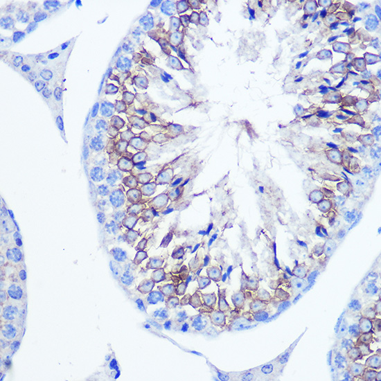 | Immunohistochemistry analysis of paraffin-embedded Mouse testis using [KO Validated] Fascin/FSCN1 Rabbit pAb (A1904) at dilution of 1:100 (40x lens). Microwave antigen retrieval performed with 0.01M PBS Buffer (pH 7.2) prior to IHC staining. |
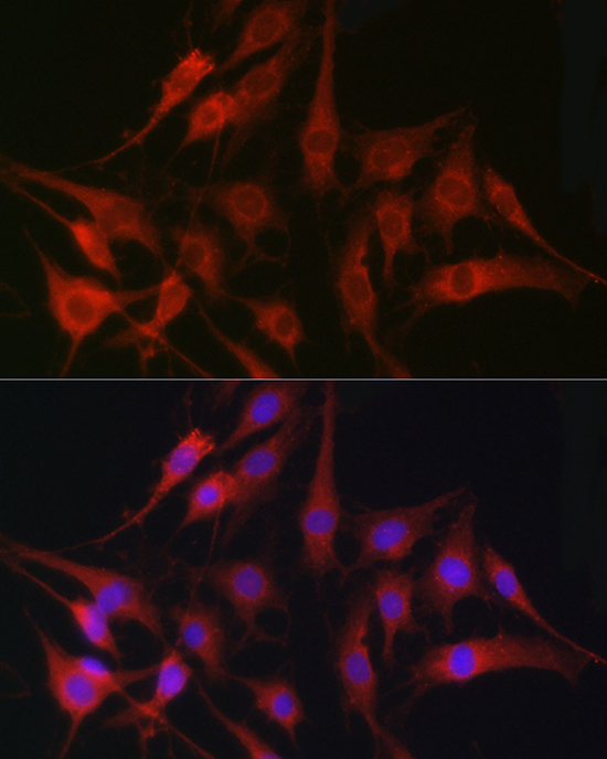 | Immunofluorescence analysis of C6 cells using [KO Validated] Fascin/Fascin/FSCN1 Rabbit pAb (A1904) at dilution of 1:100 (40x lens). Secondary antibody: Cy3-conjugated Goat anti-Rabbit IgG (H+L) (AS007) at 1:500 dilution. Blue: DAPI for nuclear staining. |
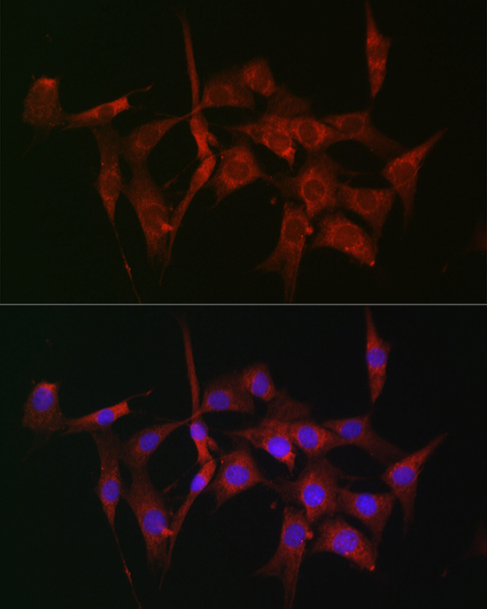 | Immunofluorescence analysis of NIH-3T3 cells using [KO Validated] Fascin/Fascin/FSCN1 Rabbit pAb (A1904) at dilution of 1:100 (40x lens). Secondary antibody: Cy3-conjugated Goat anti-Rabbit IgG (H+L) (AS007) at 1:500 dilution. Blue: DAPI for nuclear staining. |
You may also be interested in:

