Your shopping cart is empty!
| Reactivity: | Human |
| Applications: | WB, IHC-P, IF/ICC, IP, ELISA |
| Host Species: | Rabbit |
| Isotype: | IgG |
| Clonality: | Monoclonal antibody |
| Gene Name: | histone deacetylase 8 |
| Gene Symbol: | HDAC8 |
| Synonyms: | HD8; WTS; RPD3; CDA07; CDLS5; KDAC8; MRXS6; HDACL1; C8 |
| Gene ID: | 55869 |
| UniProt ID: | Q9BY41 |
| Clone ID: | 1O2Y5 |
| Immunogen: | A synthetic peptide corresponding to a sequence within amino acids 1-100 of human HDAC8 (Q9BY41). |
| Dilution: | WB 1:500-1:1000; IF/IC 1:50-1:200 |
| Purification Method: | Affinity purification |
| Concentration: | 0.3 mg/ml |
| Buffer: | PBS with 0.02% sodium azide, 0.05% BSA, 50% glycerol, pH7.3. |
| Storage: | Store at -20°C. Avoid freeze / thaw cycles. |
| Documents: | Manual-HDAC8 monoclonal antibody |
Background
Histones play a critical role in transcriptional regulation, cell cycle progression, and developmental events. Histone acetylation/deacetylation alters chromosome structure and affects transcription factor access to DNA. The protein encoded by this gene belongs to class I of the histone deacetylase family. It catalyzes the deacetylation of lysine residues in the histone N-terminal tails and represses transcription in large multiprotein complexes with transcriptional co-repressors. Multiple transcript variants encoding different isoforms have been found for this gene.
Images
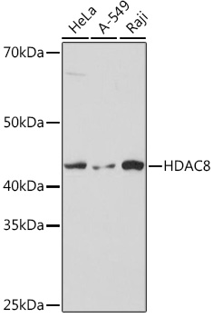 | Western blot analysis of various lysates using HDAC8 Rabbit mAb (A8865) at 1:1000 dilution. Secondary antibody: HRP-conjugated Goat anti-Rabbit IgG (H+L) (AS014) at 1:10000 dilution. Lysates/proteins: 25μg per lane. Blocking buffer: 3% nonfat dry milk in TBST. Detection: ECL Basic Kit (RM00020). Exposure time: 1s. |
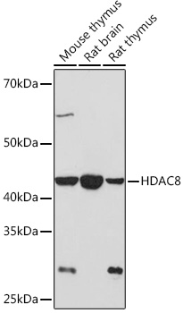 | Western blot analysis of various lysates using HDAC8 Rabbit mAb (A8865) at 1:1000 dilution. Secondary antibody: HRP-conjugated Goat anti-Rabbit IgG (H+L) (AS014) at 1:10000 dilution. Lysates/proteins: 25μg per lane. Blocking buffer: 3% nonfat dry milk in TBST. Detection: ECL Basic Kit (RM00020). Exposure time: 3min. |
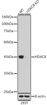 | Western blot analysis of lysates from wild type (WT) and HDAC8 knockout (KO) 293T cells, using [KO Validated] HDAC8 Rabbit mAb (A8865) at 1:1000 dilution. Secondary antibody: HRP-conjugated Goat anti-Rabbit IgG (H+L) (AS014) at 1:10000 dilution. Lysates/proteins: 25μg per lane. Blocking buffer: 3% nonfat dry milk in TBST. Detection: ECL Basic Kit (RM00020). Exposure time: 1s. |
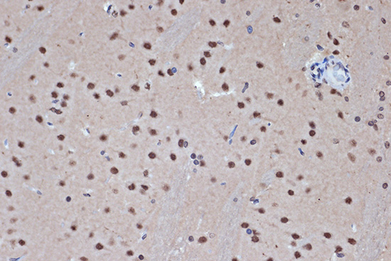 | Immunohistochemistry analysis of paraffin-embedded Rat brain using HDAC8 Rabbit mAb (A8865) at dilution of 1:100 (40x lens). Microwave antigen retrieval performed with 0.01M Tris/EDTA Buffer (pH 9.0) prior to IHC staining. |
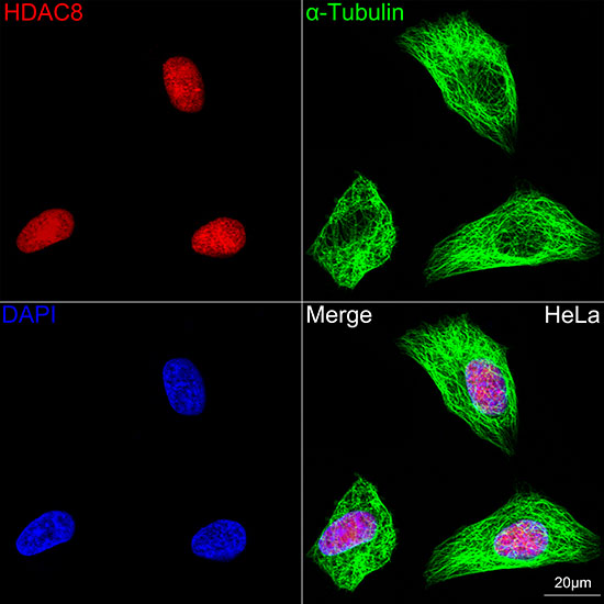 | Confocal imaging of HeLa cells using [KO Validated] HDAC8 Rabbit mAb (A8865,dilution 1:100)(Red). The cells were counterstained with α-Tubulin Mouse mAb (AC012,dilution 1:400) (Green). DAPI was used for nuclear staining (blue). Objective: 100x. |
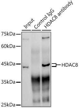 | Immunoprecipitation analysis of 300 μg extracts from HeLa cells using 3 μg HDAC8 antibody (A8865). Western blot was performed from the immunoprecipitate using HDAC8 antibody (A8865) at a dilution of 1:1000. |
You may also be interested in:

