Your shopping cart is empty!
KO-Validated HIF1AN/FIH-1 Rabbit mAb (20 μl)
| Reactivity: | Human |
| Applications: | WB, IHC-P, IF/ICC, ELISA |
| Host Species: | Rabbit |
| Isotype: | IgG |
| Clonality: | Monoclonal antibody |
| Gene Name: | hypoxia inducible factor 1 subunit alpha inhibitor |
| Gene Symbol: | HIF1AN |
| Synonyms: | FIH1 |
| Gene ID: | 55662 |
| UniProt ID: | Q9NWT6 |
| Immunogen: | Recombinant fusion protein containing a sequence corresponding to amino acids 1-349 of human HIF1AN/FIH1 (NP_060372.2). |
| Dilution: | WB 1:500-1:1000; IHC 1:50-1:200 |
| Purification Method: | Affinity purification |
| Concentration: | 1.64 mg/ml |
| Buffer: | PBS with 0.09% Sodium azide, 0.05% BSA, 50% glycerol, pH7.3. |
| Storage: | Store at -20°C. Avoid freeze / thaw cycles. |
| Documents: | Manual-HIF1AN monoclonal antibody |
Background
Enables several functions, including 2-oxoglutarate-dependent dioxygenase activity; NF-kappaB binding activity; and transition metal ion binding activity. Involved in several processes, including negative regulation of Notch signaling pathway; negative regulation of transcription from RNA polymerase II promoter in response to hypoxia; and protein hydroxylation. Located in cytosol; nucleoplasm; and perinuclear region of cytoplasm. Colocalizes with nucleus.
Images
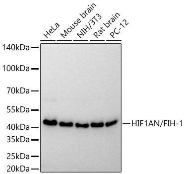 | Western blot analysis of various lysates using [KO Validated] HIF1AN/FIH-1 Rabbit mAb (A27299) at 1:16000 dilution incubated overnight at 4℃. Secondary antibody: HRP-conjugated Goat anti-Rabbit IgG (H+L) (AS014) at 1:10000 dilution. Lysates/proteins: 25 μg per lane. Blocking buffer: 3% nonfat dry milk in TBST. Detection: ECL Basic Kit (RM00020). Exposure time: 45s. |
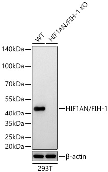 | Western blot analysis of lysates from wild type (WT) and HIF1AN/FIH-1 knockout (KO) 293T cells using [KO Validated] HIF1AN/FIH-1 Rabbit mAb (A27299) at 1:16000 dilution incubated overnight at 4℃. Secondary antibody: HRP-conjugated Goat anti-Rabbit IgG (H+L) (AS014) at 1:10000 dilution. Lysates/proteins: 25 μg per lane. Blocking buffer: 3% nonfat dry milk in TBST. Detection: ECL Basic Kit (RM00020). Exposure time: 45s. |
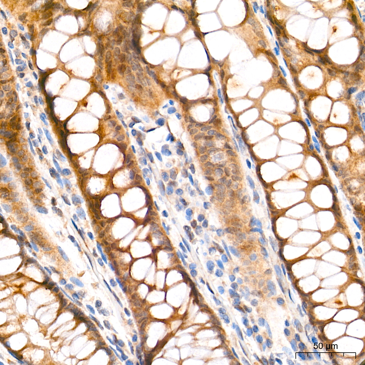 | Immunohistochemistry analysis of paraffin-embedded Human colon tissue using [KO Validated] HIF1AN/FIH-1 Rabbit mAb (A27299) at a dilution of 1:200 (40x lens). High pressure antigen retrieval performed with 0.01M Tris-EDTA Buffer (pH 9.0) prior to IHC staining. |
 | Immunohistochemistry analysis of paraffin-embedded Human esophagus tissue using [KO Validated] HIF1AN/FIH-1 Rabbit mAb (A27299) at a dilution of 1:200 (40x lens). High pressure antigen retrieval performed with 0.01M Tris-EDTA Buffer (pH 9.0) prior to IHC staining. |
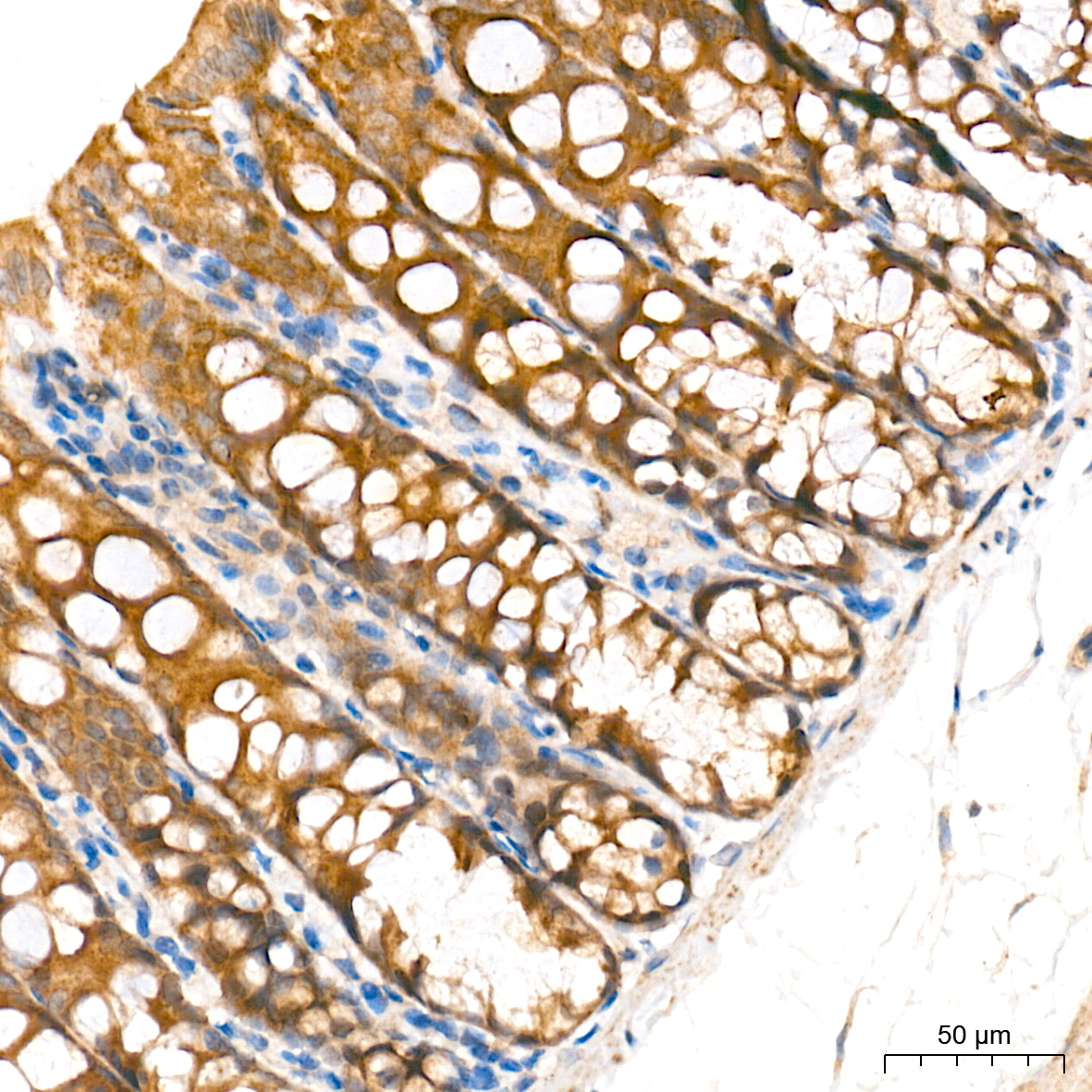 | Immunohistochemistry analysis of paraffin-embedded Mouse colon tissue using [KO Validated] HIF1AN/FIH-1 Rabbit mAb (A27299) at a dilution of 1:200 (40x lens). High pressure antigen retrieval performed with 0.01M Tris-EDTA Buffer (pH 9.0) prior to IHC staining. |
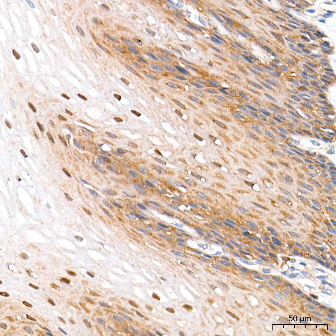
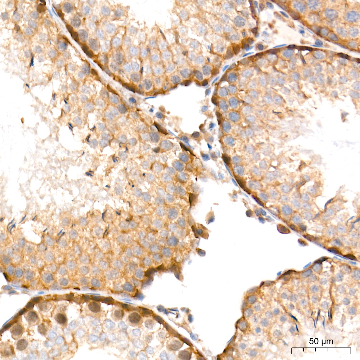 | Immunohistochemistry analysis of paraffin-embedded Rat testis tissue using [KO Validated] HIF1AN/FIH-1 Rabbit mAb (A27299) at a dilution of 1:200 (40x lens). High pressure antigen retrieval performed with 0.01M Tris-EDTA Buffer (pH 9.0) prior to IHC staining. |
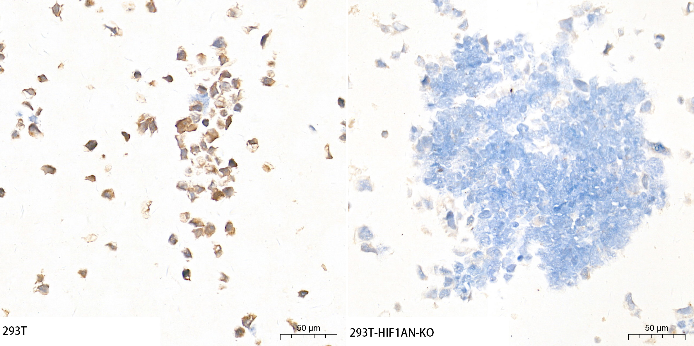 | Immunohistochemistry analysis of paraffin-embedded 293T and 293T-HIF1AN-KO cells using [KO Validated] HIF1AN/FIH-1 Rabbit mAb (A27299) at a dilution of 1:200 (40x lens). High pressure antigen retrieval performed with 0.01M Tris-EDTA Buffer (pH 9.0) prior to IHC staining. |
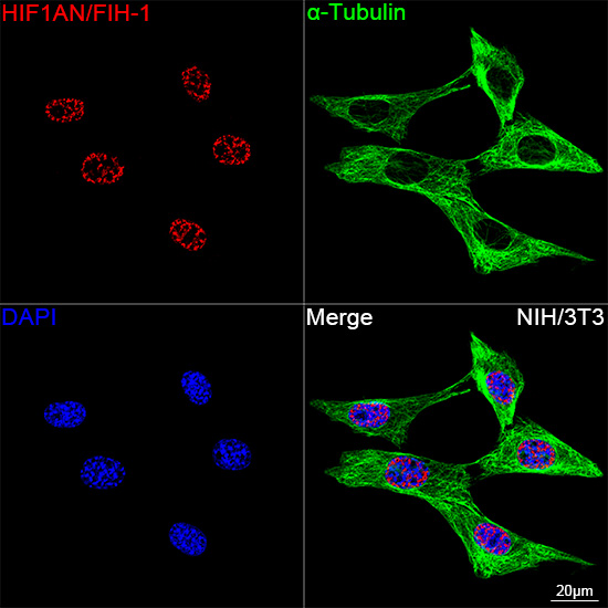 | Confocal imaging of NIH/3T3 cells using [KO Validated] HIF1AN/FIH-1 Rabbit mAb (A27299, dilution 1:200) followed by a further incubation with Cy3 Goat Anti-Rabbit IgG (H+L) (AS007, dilution 1:500) (Red). The cells were counterstained with α-Tubulin Mouse mAb (AC012, dilution 1:400) followed by incubation with ABflo® 488-conjugated Goat Anti-Mouse IgG (H+L) Ab (AS076, dilution 1:500) (Green). DAPI was used for nuclear staining (Blue). Objective: 100x. |
You may also be interested in:

