Your shopping cart is empty!
")
KO-Validated Lamin A/C Mouse mAb (20 μl)
| Reactivity: | Human, Mouse, Rat |
| Applications: | WB, IHC, IF/IC, ELISA |
| Host Species: | Mouse |
| Isotype: | IgG |
| Clonality: | Monoclonal antibody |
| Gene Name: | lamin A/C |
| Gene Symbol: | LMNA |
| Synonyms: | FPL; IDC; LFP; CDDC; EMD2; FPLD; HGPS; LDP1; LMN1; LMNC; MADA; PRO1; CDCD1; CMD1A; FPLD2; LMNL1; CMT2B1; LGMD1B |
| Gene ID: | 4000 |
| UniProt ID: | P02545 |
| Immunogen: | Recombinant fusion protein containing a sequence corresponding to amino acids 403-572 of human Lamin A/C (NP_733821.1). |
| Dilution: | WB 1:5000-1:20000; IHC 1:2000-1:20000; IF/IC 1:200-1:800 |
| Purification Method: | Affinity purification |
| Concentration: | 1.05mg/ml |
| Buffer: | PBS with 0.09% Sodium azide, 0.05% BSA, 50% glycerol, pH7.3. |
| Storage: | Store at -20°C. Avoid freeze / thaw cycles. |
| Documents: | Manual-LMNA monoclonal antibody |
Background
The protein encoded by this gene is part of the nuclear lamina, a two-dimensional matrix of proteins located next to the inner nuclear membrane. The lamin family of proteins make up the matrix and are highly conserved in evolution. During mitosis, the lamina matrix is reversibly disassembled as the lamin proteins are phosphorylated. Lamin proteins are thought to be involved in nuclear stability, chromatin structure and gene expression. Vertebrate lamins consist of two types, A and B. Alternative splicing results in multiple transcript variants. Mutations in this gene lead to several diseases: Emery-Dreifuss muscular dystrophy, familial partial lipodystrophy, limb girdle muscular dystrophy, dilated cardiomyopathy, Charcot-Marie-Tooth disease, and Hutchinson-Gilford progeria syndrome.
Images
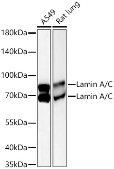 | Western blot analysis of various lysates using [KO Validated] Lamin A/C Mouse mAb (A26876) at 1:10000 dilution incubated overnight at 4℃. Secondary antibody: HRP Goat Anti-Mouse IgG (H+L) antibody (AS003) at 1:10000 dilution. Lysates/proteins: 25 μg per lane. Blocking buffer: 3% nonfat dry milk in TBST. Detection: ECL Basic Kit (RM00020). Exposure time: 45s. |
 | Western blot analysis of lysates from wild type (WT) and Lamin A/C knockout (KO) 293T cells using [KO Validated] Lamin A/C Mouse mAb (A26876) at 1:10000 dilution incubated overnight at 4℃. Secondary antibody: HRP Goat Anti-Mouse IgG (H+L) antibody (AS003) at 1:10000 dilution. Lysates/proteins: 25 μg per lane. Blocking buffer: 3% nonfat dry milk in TBST. Detection: ECL Basic Kit (RM00020). Exposure time: 90s. |
 | Immunohistochemistry analysis of paraffin-embedded Human colon carcinoma tissue using [KO Validated] Lamin A/C Mouse mAb (A26876) at a dilution of 1:10000 (40x lens). High pressure antigen retrieval performed with 0.01M Tris-EDTA Buffer(pH 9.0) prior to IHC staining. |
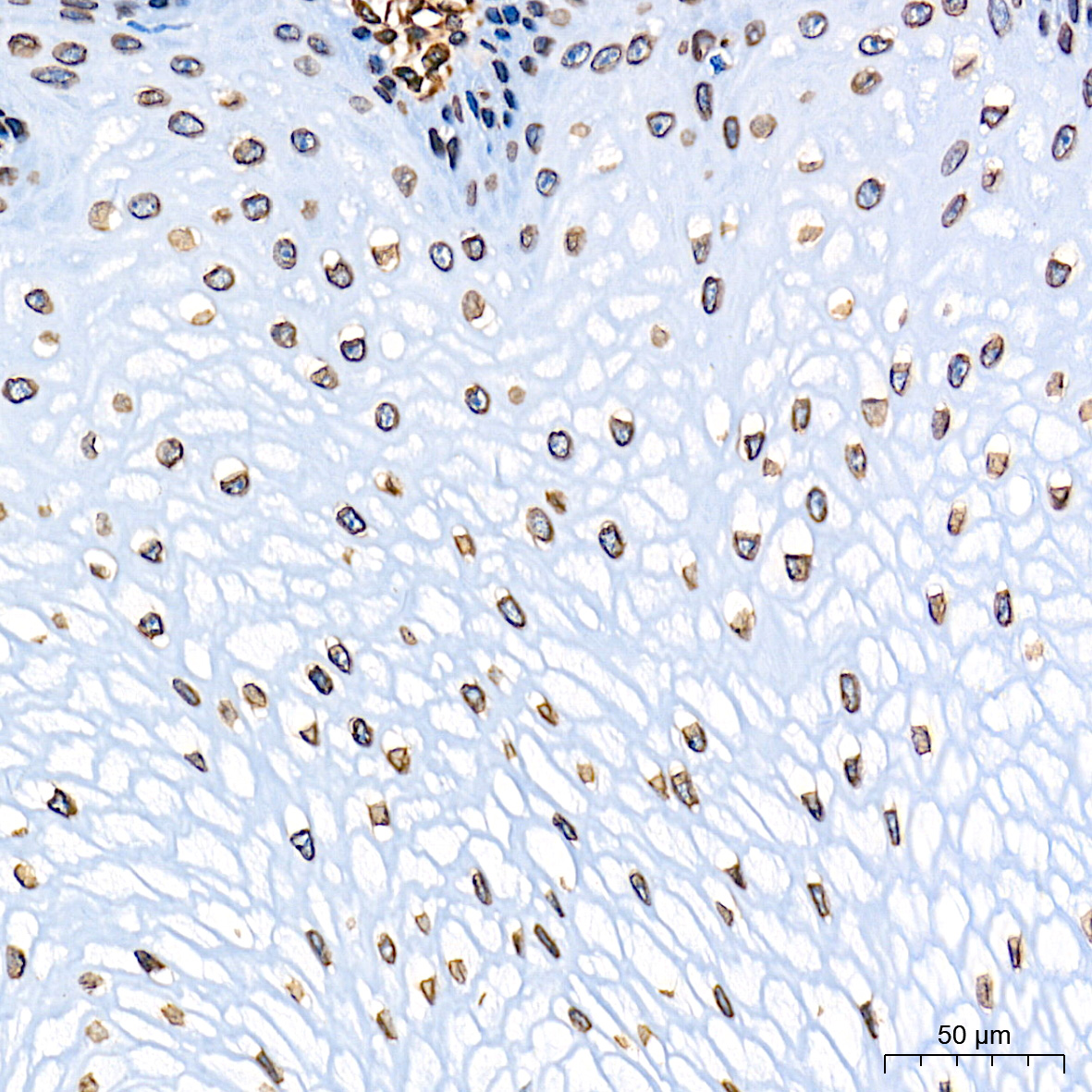 | Immunohistochemistry analysis of paraffin-embedded Human esophagus tissue using [KO Validated] Lamin A/C Mouse mAb (A26876) at a dilution of 1:10000 (40x lens). High pressure antigen retrieval performed with 0.01M Tris-EDTA Buffer(pH 9.0) prior to IHC staining. |
 | Immunohistochemistry analysis of paraffin-embedded Mouse brain tissue using [KO Validated] Lamin A/C Mouse mAb (A26876) at a dilution of 1:10000 (40x lens). High pressure antigen retrieval performed with 0.01M Tris-EDTA Buffer(pH 9.0) prior to IHC staining. |
 | Immunohistochemistry analysis of paraffin-embedded Mouse colon tissue using [KO Validated] Lamin A/C Mouse mAb (A26876) at a dilution of 1:10000 (40x lens). High pressure antigen retrieval performed with 0.01M Tris-EDTA Buffer(pH 9.0) prior to IHC staining. |
 | Immunohistochemistry analysis of paraffin-embedded Rat kidney tissue using [KO Validated] Lamin A/C Mouse mAb (A26876) at a dilution of 1:10000 (40x lens). High pressure antigen retrieval performed with 0.01M Tris-EDTA Buffer(pH 9.0) prior to IHC staining. |
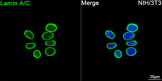 | Confocal imaging of NIH/3T3 cells using [KO Validated] Lamin A/C Mouse mAb (A26876, dilution 1:200) followed by a further incubation with ABflo® 488-conjugated Goat Anti-Mouse IgG (H+L) Ab (AS076, dilution 1:500) (Green). DAPI was used for nuclear staining (Blue). Objective: 100x. |
You may also be interested in:

