Your shopping cart is empty!
")
| Reactivity: | Human, Mouse, Rat |
| Applications: | WB, IF/IC, ELISA |
| Host Species: | Rabbit |
| Isotype: | IgG |
| Clonality: | Polyclonal antibody |
| Gene Name: | mitogen-activated protein kinase 1 |
| Gene Symbol: | MAPK1 |
| Synonyms: | ERK; p38; p40; p41; ERK2; ERT1; NS13; ERK-2; MAPK2; PRKM1; PRKM2; P42MAPK; p41mapk; p42-MAPK; K2 |
| Gene ID: | 5594 |
| UniProt ID: | P28482 |
| Immunogen: | A synthetic peptide corresponding to a sequence within amino acids 200-300 of human ERK2 (NP_002736.3). |
| Dilution: | WB 1:500-1:1000; IF/IC 1:50-1:200 |
| Purification Method: | Affinity purification |
| Concentration: | 2.29 mg/ml |
| Buffer: | PBS with 0.09% Sodium azide, 50% glycerol, pH7.3. |
| Storage: | Store at -20°C. Avoid freeze / thaw cycles. |
| Documents: | Manual-MAPK1 polyclonal antibody |
Background
This gene encodes a member of the MAP kinase family. MAP kinases, also known as extracellular signal-regulated kinases (ERKs), act as an integration point for multiple biochemical signals, and are involved in a wide variety of cellular processes such as proliferation, differentiation, transcription regulation and development. The activation of this kinase requires its phosphorylation by upstream kinases. Upon activation, this kinase translocates to the nucleus of the stimulated cells, where it phosphorylates nuclear targets. One study also suggests that this protein acts as a transcriptional repressor independent of its kinase activity. The encoded protein has been identified as a moonlighting protein based on its ability to perform mechanistically distinct functions. Two alternatively spliced transcript variants encoding the same protein, but differing in the UTRs, have been reported for this gene.
Images
 | Western blot analysis of various lysates using [KO Validated] ERK2 Rabbit pAb (A0229) at 1:1000 dilution. Secondary antibody: HRP-conjugated Goat anti-Rabbit IgG (H+L) (AS014) at 1:10000 dilution. Lysates/proteins: 25μg per lane. Blocking buffer: 3% nonfat dry milk in TBST. Detection: ECL Basic Kit (RM00020). Exposure time: 10s. |
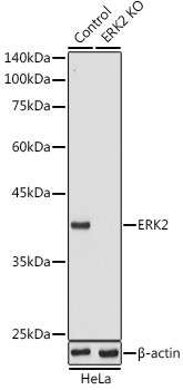 | Western blot analysis of lysates from wild type (WT) and ERK2 knockout (KO) HeLa cells, using [KO Validated] ERK2 Rabbit pAb (A0229) at 1:1000 dilution. Secondary antibody: HRP-conjugated Goat anti-Rabbit IgG (H+L) (AS014) at 1:10000 dilution. Lysates/proteins: 25μg per lane. Blocking buffer: 3% nonfat dry milk in TBST. Detection: ECL Basic Kit (RM00020). Exposure time: 10s. |
You may also be interested in:

