Your shopping cart is empty!
| Reactivity: | Human |
| Applications: | WB, IHC-P, IF/ICC, ELISA |
| Host Species: | Rabbit |
| Isotype: | IgG |
| Clonality: | Polyclonal antibody |
| Gene Name: | nuclear receptor binding factor 2 |
| Gene Symbol: | NRBF2 |
| Synonyms: | COPR; COPR1; COPR2; NRBF-2; F2 |
| Gene ID: | 29982 |
| UniProt ID: | Q96F24 |
| Immunogen: | Recombinant fusion protein containing a sequence corresponding to amino acids 1-287 of human NRBF2 (NP_110386.2). |
| Dilution: | WB 1:500-1:2000 |
| Purification Method: | Affinity purification |
| Concentration: | 2.38 mg/ml |
| Buffer: | PBS with 0.09% Sodium azide, 50% glycerol, pH7.3. |
| Storage: | Store at -20°C. Avoid freeze / thaw cycles. |
| Documents: | Manual-NRBF2 polyclonal antibody |
Background
Involved in autophagy. Located in cytoplasm. Colocalizes with phosphatidylinositol 3-kinase complex, class III.
Images
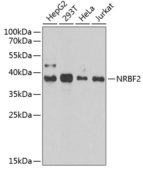 | Western blot analysis of various lysates using NRBF2 Rabbit pAb (A6462) at 1:1000 dilution. Secondary antibody: HRP-conjugated Goat anti-Rabbit IgG (H+L) (AS014) at 1:10000 dilution. Lysates/proteins: 25μg per lane. Blocking buffer: 3% nonfat dry milk in TBST. Detection: ECL Enhanced Kit (RM00021). Exposure time: 90s. |
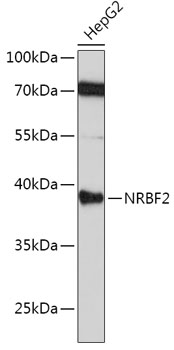 | Western blot analysis of lysates from HepG2 cells, using [KO Validated] NRBF2 Rabbit pAb (A6462) at 1:1000 dilution. Secondary antibody: HRP-conjugated Goat anti-Rabbit IgG (H+L) (AS014) at 1:10000 dilution. Lysates/proteins: 25μg per lane. Blocking buffer: 3% nonfat dry milk in TBST. Detection: ECL Basic Kit (RM00020). Exposure time: 3min. |
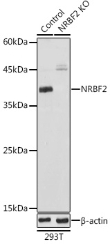 | Western blot analysis of lysates from wild type (WT) and NRBF2 knockout (KO) 293T cells, using [KO Validated] NRBF2 Rabbit pAb (A6462) at 1:1000 dilution. Secondary antibody: HRP-conjugated Goat anti-Rabbit IgG (H+L) (AS014) at 1:10000 dilution. Lysates/proteins: 25μg per lane. Blocking buffer: 3% nonfat dry milk in TBST. Detection: ECL Basic Kit (RM00020). Exposure time: 10s. |
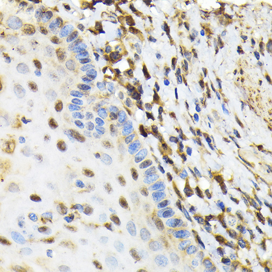 | Immunohistochemistry analysis of paraffin-embedded Human esophageal using [KO Validated] NRBF2 Rabbit pAb (A6462) at dilution of 1:100 (40x lens). Microwave antigen retrieval performed with 0.01M Tris/EDTA Buffer (pH 9.0) prior to IHC staining. |
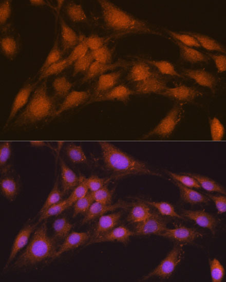 | Immunofluorescence analysis of C6 cells using [KO Validated] NRBF2 Rabbit pAb (A6462) at dilution of 1:100. Secondary antibody: Cy3-conjugated Goat anti-Rabbit IgG (H+L) (AS007) at 1:500 dilution. Blue: DAPI for nuclear staining. |
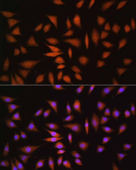 | Immunofluorescence analysis of L929 cells using [KO Validated] NRBF2 Rabbit pAb (A6462) at dilution of 1:100. Secondary antibody: Cy3-conjugated Goat anti-Rabbit IgG (H+L) (AS007) at 1:500 dilution. Blue: DAPI for nuclear staining. |
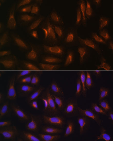 | Immunofluorescence analysis of U-2 OS cells using [KO Validated] NRBF2 Rabbit pAb (A6462) at dilution of 1:100. Secondary antibody: Cy3-conjugated Goat anti-Rabbit IgG (H+L) (AS007) at 1:500 dilution. Blue: DAPI for nuclear staining. |
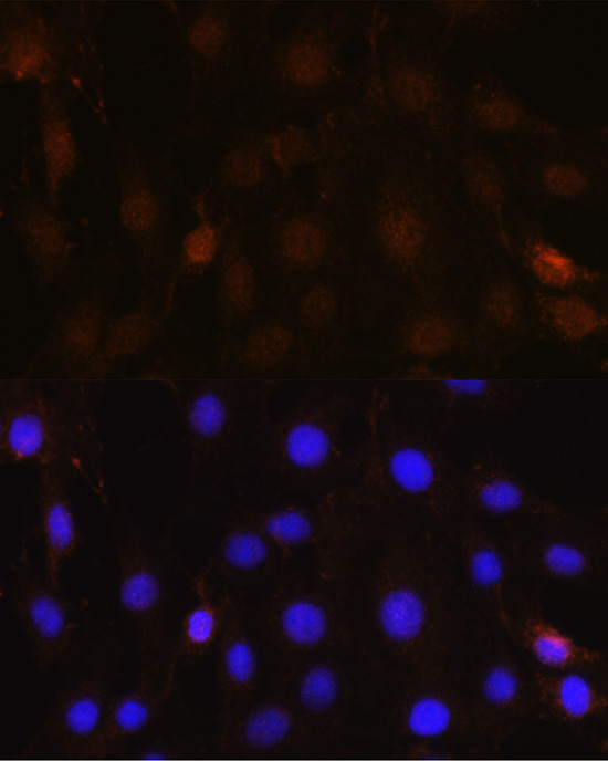 | Immunofluorescence analysis of C6 cells using NRBF2 Rabbit pAb (A6462) at dilution of 1:100 (40x lens). Secondary antibody: Cy3-conjugated Goat anti-Rabbit IgG (H+L) (AS007) at 1:500 dilution. Blue: DAPI for nuclear staining. |
You may also be interested in:

