Your shopping cart is empty!
")
KO-Validated hnRNP E1/PCBP1 Rabbit pAb (20 μl)
| Reactivity: | Human, Mouse, Rat |
| Applications: | WB, IHC, ELISA |
| Host Species: | rabbit |
| Isotype: | IgG |
| Clonality: | Polyclonal antibody |
| Gene Name: | poly(rC) binding protein 1 |
| Gene Symbol: | PCBP1 |
| Synonyms: | HNRPX; HNRPE1; hnRNP-X; HEL-S-85; hnRNP-E1; P1 |
| Gene ID: | 5093 |
| UniProt ID: | Q15365 |
| Immunogen: | A synthetic peptide corresponding to a sequence within amino acids 100-200 of human hnRNP E1/PCBP1 (NP_006187.2). |
| Dilution: | WB 1:500-1:1000; IHC 1:50-1:200 |
| Purification Method: | Affinity purification |
| Concentration: | 1.18 mg/ml |
| Buffer: | PBS with 0.05% proclin300, 50% glycerol, pH7.3. |
| Storage: | Store at -20°C. Avoid freeze / thaw cycles. |
| Documents: | Manual-PCBP1 polyclonal antibody |
Background
This intronless gene is thought to have been generated by retrotransposition of a fully processed PCBP-2 mRNA. This gene and PCBP-2 have paralogues (PCBP3 and PCBP4) which are thought to have arisen as a result of duplication events of entire genes. The protein encoded by this gene appears to be multifunctional. It along with PCBP-2 and hnRNPK corresponds to the major cellular poly(rC)-binding protein. It contains three K-homologous (KH) domains which may be involved in RNA binding. This encoded protein together with PCBP-2 also functions as translational coactivators of poliovirus RNA via a sequence-specific interaction with stem-loop IV of the IRES and promote poliovirus RNA replication by binding to its 5'-terminal cloverleaf structure. It has also been implicated in translational control of the 15-lipoxygenase mRNA, human Papillomavirus type 16 L2 mRNA, and hepatitis A virus RNA. The encoded protein is also suggested to play a part in formation of a sequence-specific alpha-globin mRNP complex which is associated with alpha-globin mRNA stability.
Images
 | Western blot analysis of various lysates using [KO Validated] hnRNP E1/PCBP1 Rabbit pAb (A21733) at 1:1000 dilution. Secondary antibody: HRP-conjugated Goat anti-Rabbit IgG (H+L) (AS014) at 1:10000 dilution. Lysates/proteins: 25μg per lane. Blocking buffer: 3% nonfat dry milk in TBST. Detection: ECL Basic Kit (RM00020). Exposure time: 30s. |
 | Western blot analysis of lysates from wild type(WT) and hnRNP E1/PCBP1 knockout (KO) HeLa cells, using [KO Validated] hnRNP E1/PCBP1 Rabbit pAb (A21733) at 1:1000 dilution. Secondary antibody: HRP-conjugated Goat anti-Rabbit IgG (H+L) (AS014) at 1:10000 dilution. Lysates/proteins: 25μg per lane. Blocking buffer: 3% nonfat dry milk in TBST. Detection: ECL Basic Kit (RM00020). Exposure time: 30s. |
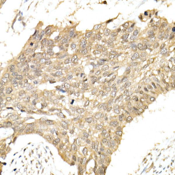 | Immunohistochemistry analysis of paraffin-embedded Human esophageal cancer using hnRNP E1/PCBP1 Rabbit pAb (A21733) at dilution of 1:100 (40x lens). High pressure antigen retrieval performed with 0.01M Citrate Bufferr (pH 6.0) prior to IHC staining. |
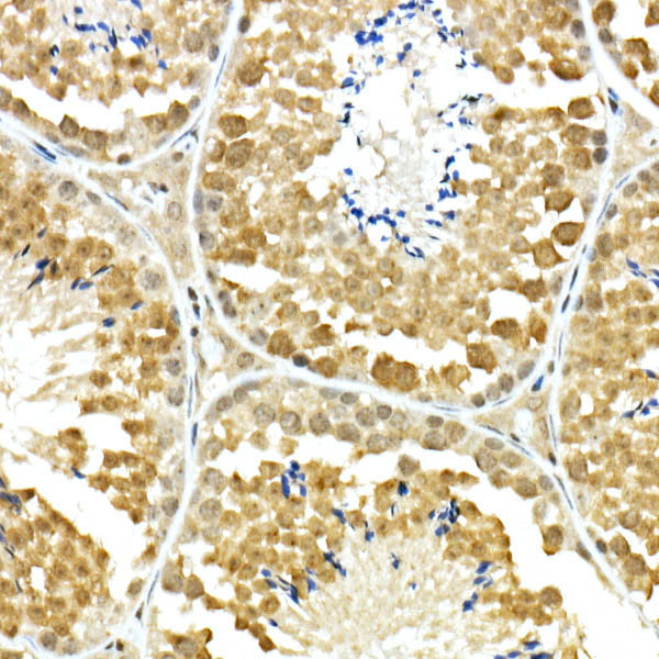 | Immunohistochemistry analysis of paraffin-embedded Mouse testis using hnRNP E1/PCBP1 Rabbit pAb (A21733) at dilution of 1:100 (40x lens). High pressure antigen retrieval performed with 0.01M Citrate Bufferr (pH 6.0) prior to IHC staining. |
 | Immunohistochemistry analysis of paraffin-embedded Rat brain using hnRNP E1/PCBP1 Rabbit pAb (A21733) at dilution of 1:100 (40x lens). High pressure antigen retrieval performed with 0.01M Citrate Bufferr (pH 6.0) prior to IHC staining. |
You may also be interested in:

