Your shopping cart is empty!
| Reactivity: | Human |
| Applications: | WB, IHC-P, IP, ELISA |
| Host Species: | Rabbit |
| Isotype: | IgG |
| Clonality: | Monoclonal antibody |
| Gene Name: | phosphorylated CTD interacting factor 1 |
| Gene Symbol: | PCIF1 |
| Synonyms: | CAPAM; hCAPAM; hPCIF1; C20orf67; PPP1R121; F1 |
| Gene ID: | 63935 |
| UniProt ID: | Q9H4Z3 |
| Clone ID: | 8X8U1 |
| Immunogen: | Recombinant fusion protein containing a sequence corresponding to amino acids 1-200 of human PCIF1(NP_071387.1). |
| Dilution: | WB 1:500-1:1000; IF/IC 1:50-1:100 |
| Purification Method: | Affinity purification |
| Concentration: | 1.76 mg/ml |
| Buffer: | PBS with 0.05% proclin300, 0.05% BSA, 50% glycerol, pH7.3. |
| Storage: | Store at -20°C. Avoid freeze / thaw cycles. |
| Documents: | Manual-PCIF1 monoclonal antibody |
Background
Enables RNA polymerase II C-terminal domain phosphoserine binding activity; S-adenosyl-L-methionine binding activity; and mRNA (2'-O-methyladenosine-N6-)-methyltransferase activity. Involved in mRNA methylation; negative regulation of translation; and positive regulation of translation. Located in intercellular bridge; microtubule cytoskeleton; and nucleoplasm.
Images
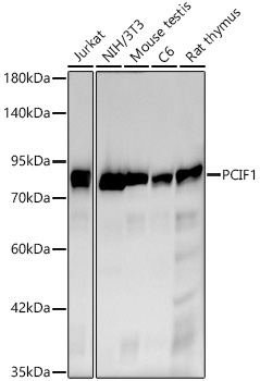 | Western blot analysis of various lysates using [KO Validated] PCIF1 Rabbit mAb (A24070) at 1:1000 dilution. Secondary antibody: HRP-conjugated Goat anti-Rabbit IgG (H+L) (AS014) at 1:10000 dilution. Lysates / proteins: 25 μg per lane. Blocking buffer: 3 % nonfat dry milk in TBST. Detection: ECL Basic Kit (RM00020). Exposure time: 45s. |
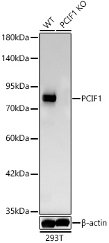 | Western blot analysis of lysates from wild type (WT) and PCIF1 knockout (KO) 293T cells (RM01804) using [KO Validated] PCIF1 Rabbit mAb (A24070) at 1:1000 dilution. Secondary antibody: HRP-conjugated Goat anti-Rabbit IgG (H+L) (AS014) at 1:10000 dilution. Lysates/proteins: 25 μg per lane. Blocking buffer: 3% nonfat dry milk in TBST. Detection: ECL Basic Kit (RM00020). Exposure time: 45s. |
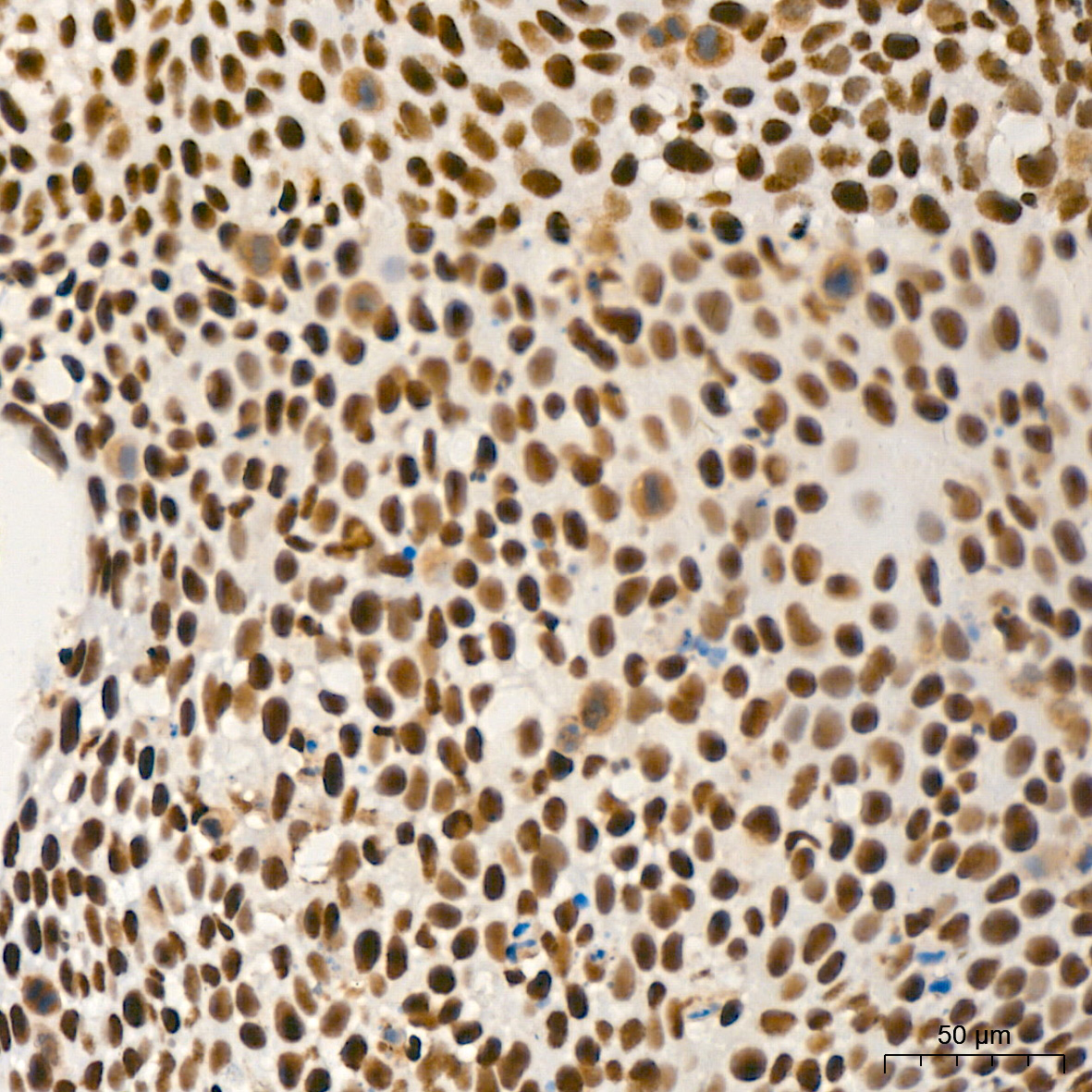 | Immunohistochemistry analysis of paraffin-embedded Human cervix cancer tissue using [KO Validated] PCIF1 Rabbit mAb (A24070) at a dilution of 1:200 (40x lens). High pressure antigen retrieval performed with 0.01M Citrate Bufferr (pH 6.0) prior to IHC staining. |
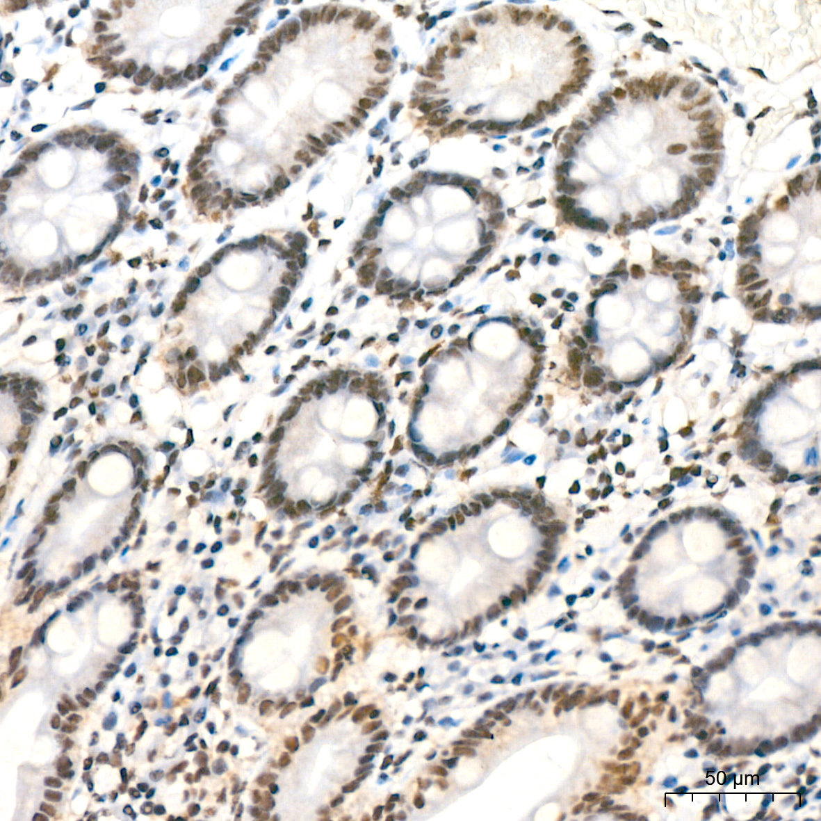 | Immunohistochemistry analysis of paraffin-embedded Human colon tissue using [KO Validated] PCIF1 Rabbit mAb (A24070) at a dilution of 1:200 (40x lens). High pressure antigen retrieval performed with 0.01M Citrate Bufferr (pH 6.0) prior to IHC staining. |
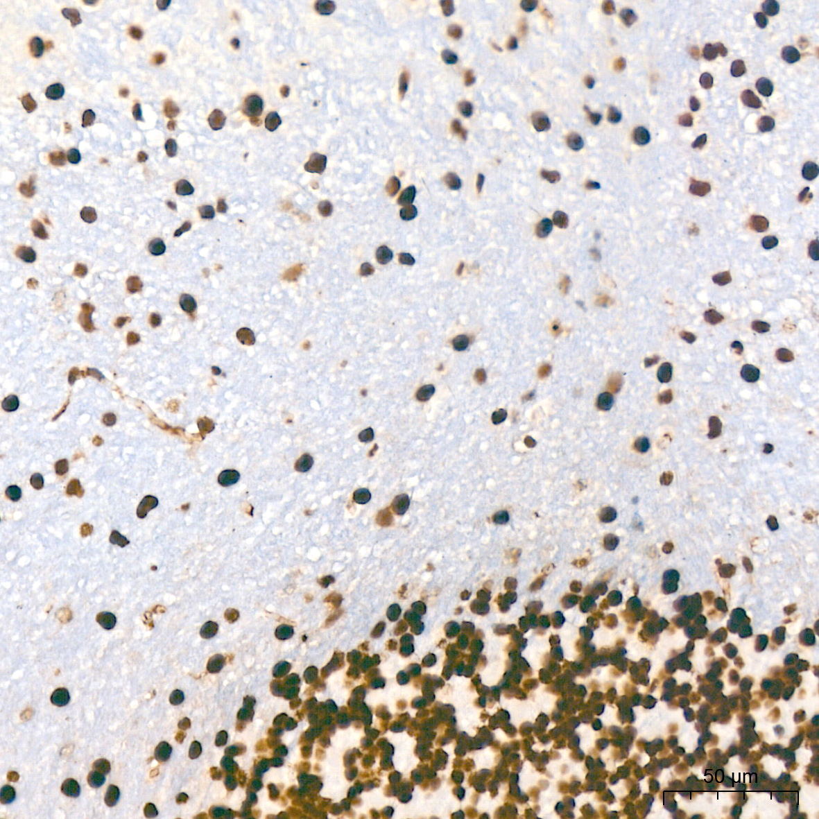 | Immunohistochemistry analysis of paraffin-embedded Mouse brain tissue using [KO Validated] PCIF1 Rabbit mAb (A24070) at a dilution of 1:200 (40x lens). High pressure antigen retrieval performed with 0.01M Citrate Bufferr (pH 6.0) prior to IHC staining. |
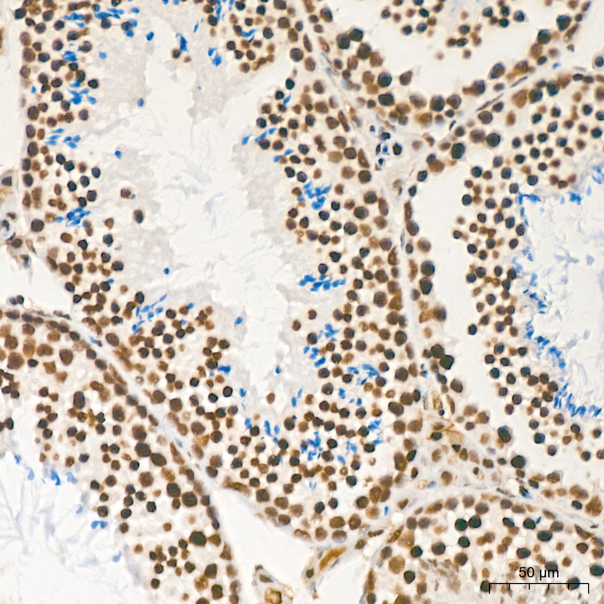 | Immunohistochemistry analysis of paraffin-embedded Mouse testis tissue using [KO Validated] PCIF1 Rabbit mAb (A24070) at a dilution of 1:200 (40x lens). High pressure antigen retrieval performed with 0.01M Citrate Bufferr (pH 6.0) prior to IHC staining. |
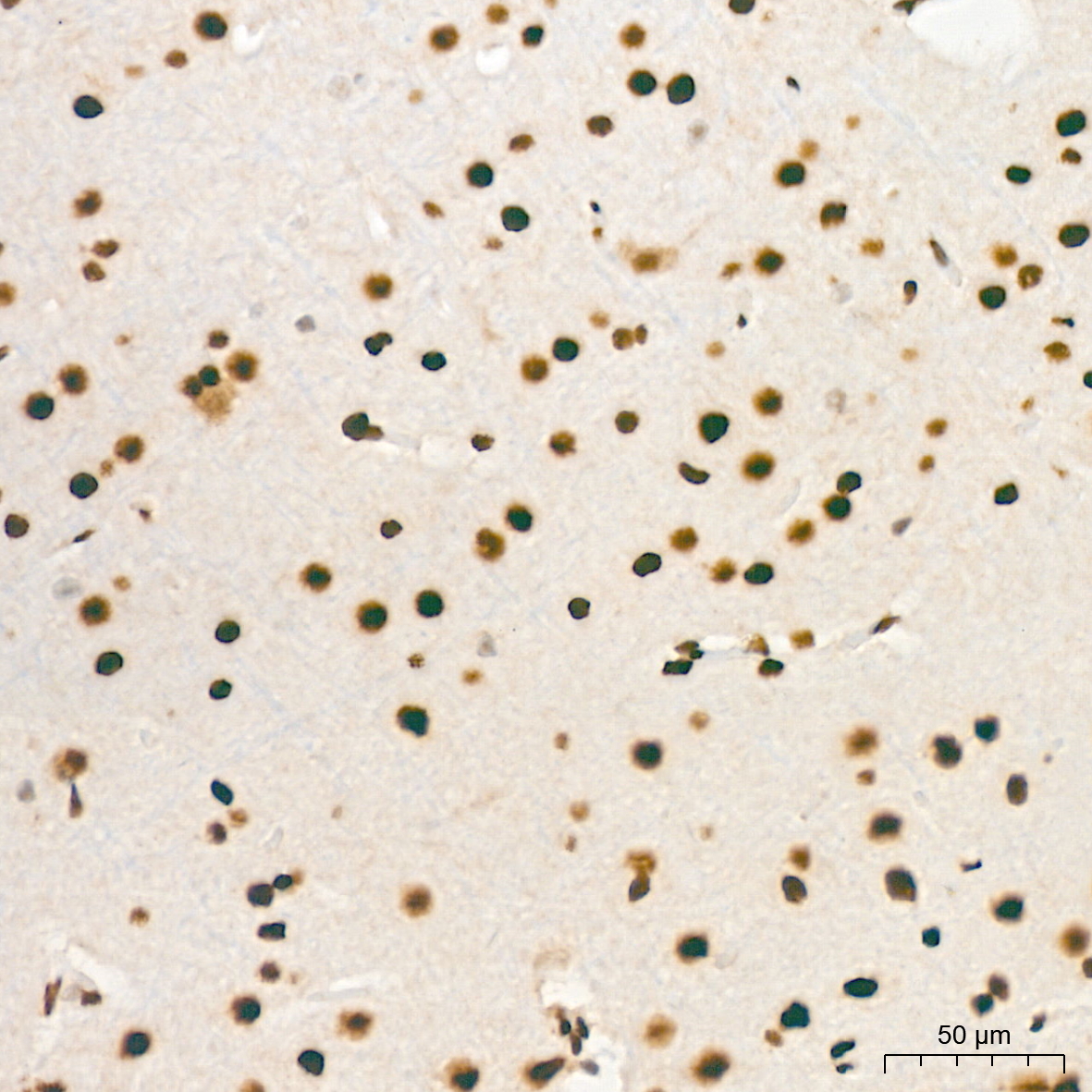 | Immunohistochemistry analysis of paraffin-embedded Rat brain tissue using [KO Validated] PCIF1 Rabbit mAb (A24070) at a dilution of 1:200 (40x lens). High pressure antigen retrieval performed with 0.01M Citrate Bufferr (pH 6.0) prior to IHC staining. |
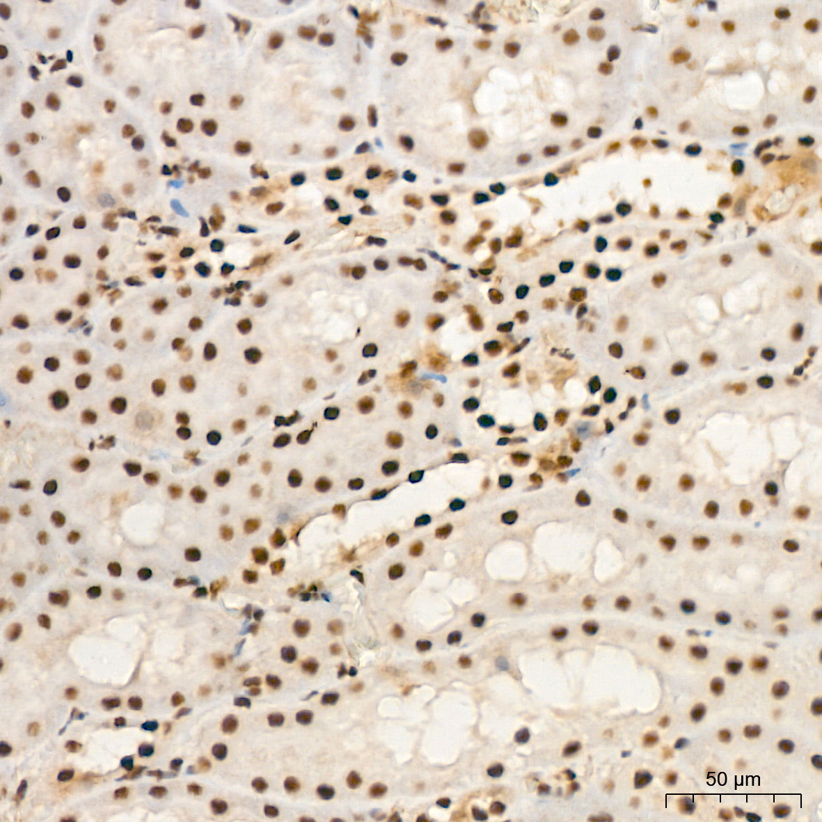 | Immunohistochemistry analysis of paraffin-embedded Rat kidney tissue using [KO Validated] PCIF1 Rabbit mAb (A24070) at a dilution of 1:200 (40x lens). High pressure antigen retrieval performed with 0.01M Citrate Bufferr (pH 6.0) prior to IHC staining. |
You may also be interested in:

