Your shopping cart is empty!
| Reactivity: | Human |
| Applications: | WB, IHC-P, ELISA |
| Host Species: | Rabbit |
| Isotype: | IgG |
| Clonality: | Polyclonal antibody |
| Gene Name: | programmed cell death 4 |
| Gene Symbol: | PDCD4 |
| Synonyms: | H731; D4 |
| Gene ID: | 27250 |
| UniProt ID: | Q53EL6 |
| Immunogen: | Recombinant fusion protein containing a sequence corresponding to amino acids 1-260 of human PDCD4 (NP_055271.2). |
| Dilution: | WB 1:2000-1:8000 |
| Purification Method: | Affinity purification |
| Concentration: | 0.3 mg/ml |
| Buffer: | PBS with 0.02% sodium azide, 50% glycerol ,pH7.3. |
| Storage: | Store at -20°C. Avoid freeze / thaw cycles. |
| Documents: | Manual-PDCD4 polyclonal antibody |
Background
This gene is a tumor suppressor and encodes a protein that binds to the eukaryotic translation initiation factor 4A1 and inhibits its function by preventing RNA binding. Alternative splicing results in multiple transcript variants.
Images
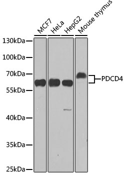 | Western blot analysis of various lysates using [KO Validated] PDCD4 Rabbit pAb (A2570) at 1:1000 dilution. Secondary antibody: HRP-conjugated Goat anti-Rabbit IgG (H+L) (AS014) at 1:10000 dilution. Lysates/proteins: 25μg per lane. Blocking buffer: 3% nonfat dry milk in TBST. Detection: ECL Enhanced Kit (RM00021). Exposure time: 90s. |
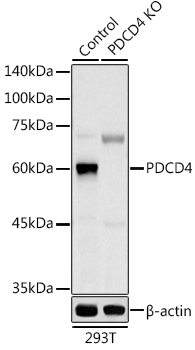 | Western blot analysis of lysates from wild type (WT) and PDCD4 knockout (KO) 293T cells, using [KO Validated] PDCD4 Rabbit pAb (A2570) at 1:1000 dilution. Secondary antibody: HRP-conjugated Goat anti-Rabbit IgG (H+L) (AS014) at 1:10000 dilution. Lysates/proteins: 25μg per lane. Blocking buffer: 3% nonfat dry milk in TBST. Detection: ECL Basic Kit (RM00020). Exposure time: 180s. |
 | Western blot analysis of lysates from Hep G2 cells using PDCD4 Rabbit pAb (A2570) at 1:1000 dilution. Secondary antibody: HRP-conjugated Goat anti-Rabbit IgG (H+L) (AS014) at 1:10000 dilution. Lysates/proteins: 25 μg per lane. Blocking buffer: 3% nonfat dry milk in TBST. Detection: ECL Basic Kit (RM00020). Exposure time: 30s. |
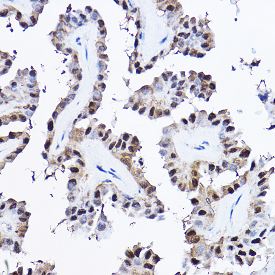
 | Immunohistochemistry analysis of paraffin-embedded Human thyroid cancer using [KO Validated] PDCD4 Rabbit pAb (A2570) at dilution of 1:20 (40x lens). High pressure antigen retrieval performed with 0.01M Citrate Bufferr (pH 6.0) prior to IHC staining. |
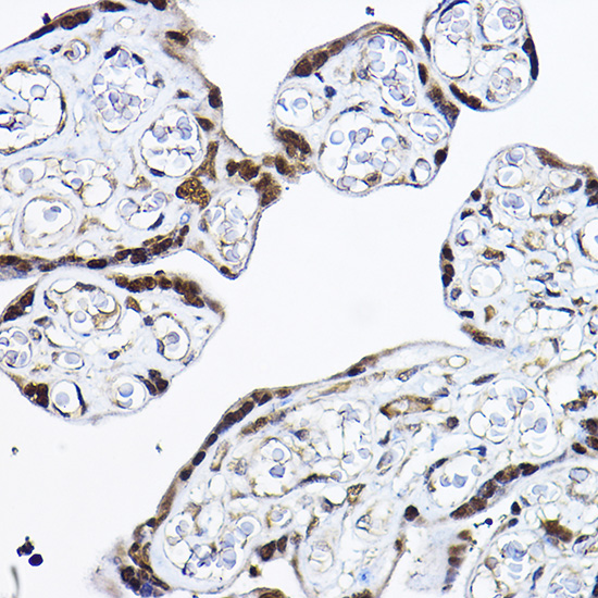
 | Immunohistochemistry analysis of paraffin-embedded Human placenta using [KO Validated] PDCD4 Rabbit pAb (A2570) at dilution of 1:20 (40x lens). High pressure antigen retrieval performed with 0.01M Citrate Bufferr (pH 6.0) prior to IHC staining. |
You may also be interested in:

