Your shopping cart is empty!
 Mouse mAb (20 μl)")
KO-Validated PKA C-alpha (PRKACA) Mouse mAb (20 μl)
| Reactivity: | Human, Mouse, Rat |
| Applications: | WB, IF/IC, ELISA |
| Host Species: | Mouse |
| Isotype: | IgG2B,Kappa |
| Clonality: | Monoclonal antibody |
| Gene Name: | protein kinase cAMP-activated catalytic subunit alpha |
| Gene Symbol: | PRKACA |
| Synonyms: | CAFD1; PKACA; PPNAD4; A) |
| Gene ID: | 5566 |
| UniProt ID: | P17612 |
| Clone ID: | 8H1Q6 |
| Immunogen: | Recombinant protein of human [KD Validated] PKA C-alpha (PRKACA). |
| Dilution: | WB 1:10000-1:40000; IF/IC 1:50-1:200 |
| Purification Method: | Affinity purification |
| Concentration: | 0.5 mg/ml |
| Buffer: | PBS with 0.02% sodium azide, 50% glycerol ,pH7.3. |
| Storage: | Store at -20°C. Avoid freeze / thaw cycles. |
| Documents: | Manual-PRKACA monoclonal antibody |
Background
This gene encodes one of the catalytic subunits of protein kinase A, which exists as a tetrameric holoenzyme with two regulatory subunits and two catalytic subunits, in its inactive form. cAMP causes the dissociation of the inactive holoenzyme into a dimer of regulatory subunits bound to four cAMP and two free monomeric catalytic subunits. Four different regulatory subunits and three catalytic subunits have been identified in humans. cAMP-dependent phosphorylation of proteins by protein kinase A is important to many cellular processes, including differentiation, proliferation, and apoptosis. Constitutive activation of this gene caused either by somatic mutations, or genomic duplications of regions that include this gene, have been associated with hyperplasias and adenomas of the adrenal cortex and are linked to corticotropin-independent Cushing's syndrome. Alternative splicing results in multiple transcript variants encoding different isoforms. Tissue-specific isoforms that differ at the N-terminus have been described, and these isoforms may differ in the post-translational modifications that occur at the N-terminus of some isoforms.
Images
 | Western blot analysis of various lysates, using [KO Validated] PKA C-alpha (PRKACA) Mouse mAb (A18603) at 1:10000 dilution. Secondary antibody: HRP-conjugated Goat anti-Mouse IgG (H+L) (AS003) at 1:10000 dilution. Lysates/proteins: 25μg per lane. Blocking buffer: 3% nonfat dry milk in TBST. Detection: ECL Basic Kit (RM00020). Exposure time: 20s. |
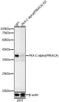 | Western blot analysis of lysates from wild type(WT) and PKA C-alpha (PRKACA) knockdown (KD) 293T(KD) cells, using [KO Validated] PKA C-alpha (PRKACA) Mouse mAb (A18603) at 1:10000 dilution. Secondary antibody: HRP-conjugated Goat anti-Mouse IgG (H+L) (AS003) at 1:10000 dilution. Lysates/proteins: 25μg per lane. Blocking buffer: 3% nonfat dry milk in TBST. Detection: ECL Basic Kit (RM00020). Exposure time: 20s. |
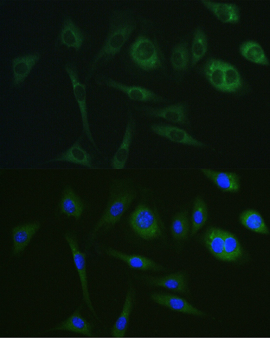 | Immunofluorescence analysis of BALB-3T3 cells using [KD Validated] PKA C-alpha (PRKACA) Mouse mAb (A18603) at dilution of 1:100 (40x lens). Blue: DAPI for nuclear staining. |
 | Immunofluorescence analysis of MCF7 cells using [KD Validated] PKA C-alpha (PRKACA) Mouse mAb (A18603) at dilution of 1:100 (40x lens). Blue: DAPI for nuclear staining. |
You may also be interested in:

