Your shopping cart is empty!
")
KO-Validated PRKAR1A Rabbit pAb (20 μl)
| Reactivity: | Human, Mouse, Rat |
| Applications: | WB, IF/IC, ELISA |
| Host Species: | Rabbit |
| Isotype: | IgG |
| Clonality: | Polyclonal antibody |
| Gene Name: | protein kinase cAMP-dependent type I regulatory subunit alpha |
| Gene Symbol: | PRKAR1A |
| Synonyms: | CAR; CNC; CNC1; PKR1; TSE1; ADOHR; PPNAD1; PRKAR1; ACRDYS1; 1A |
| Gene ID: | 5573 |
| UniProt ID: | P10644 |
| Immunogen: | Recombinant fusion protein containing a sequence corresponding to amino acids 1-250 of human PRKAR1A (NP_002725.1). |
| Dilution: | WB 1:500-1:1000; IF/IC 1:50-1:200 |
| Purification Method: | Affinity purification |
| Concentration: | 0.72 mg/ml |
| Buffer: | PBS with 0.09% Sodium azide, 50% glycerol, pH7.3. |
| Storage: | Store at -20°C. Avoid freeze / thaw cycles. |
| Documents: | Manual-PRKAR1A polyclonal antibody |
Background
cAMP is a signaling molecule important for a variety of cellular functions. cAMP exerts its effects by activating the cAMP-dependent protein kinase, which transduces the signal through phosphorylation of different target proteins. The inactive kinase holoenzyme is a tetramer composed of two regulatory and two catalytic subunits. cAMP causes the dissociation of the inactive holoenzyme into a dimer of regulatory subunits bound to four cAMP and two free monomeric catalytic subunits. Four different regulatory subunits and three catalytic subunits have been identified in humans. This gene encodes one of the regulatory subunits. This protein was found to be a tissue-specific extinguisher that down-regulates the expression of seven liver genes in hepatoma x fibroblast hybrids. Mutations in this gene cause Carney complex (CNC). This gene can fuse to the RET protooncogene by gene rearrangement and form the thyroid tumor-specific chimeric oncogene known as PTC2. A nonconventional nuclear localization sequence (NLS) has been found for this protein which suggests a role in DNA replication via the protein serving as a nuclear transport protein for the second subunit of the Replication Factor C (RFC40). Several alternatively spliced transcript variants encoding two different isoforms have been observed.
Images
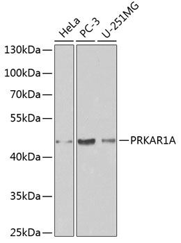 | Western blot analysis of various lysates using [KO Validated] PRKAR1A Rabbit pAb (A0906) at 1:1000 dilution. Secondary antibody: HRP-conjugated Goat anti-Rabbit IgG (H+L) (AS014) at 1:10000 dilution. Lysates/proteins: 25μg per lane. Blocking buffer: 3% nonfat dry milk in TBST. |
 | Western blot analysis of lysates from wild type (WT) and PRKAR1A knockout (KO) 293T cells, using [KO Validated] PRKAR1A Rabbit pAb (A0906) at 1:500 dilution. Secondary antibody: HRP-conjugated Goat anti-Rabbit IgG (H+L) (AS014) at 1:10000 dilution. Lysates/proteins: 25μg per lane. Blocking buffer: 3% nonfat dry milk in TBST. Detection: ECL Basic Kit (RM00020). Exposure time: 5s. |
 | Western blot analysis of lysates from wild type (WT) and PRKAR1A knockout (KO) 293T cells, using [KO Validated] PRKAR1A Rabbit pAb (A0906) at 1:1000 dilution. Secondary antibody: HRP-conjugated Goat anti-Rabbit IgG (H+L) (AS014) at 1:10000 dilution. Lysates/proteins: 25μg per lane. Blocking buffer: 3% nonfat dry milk in TBST. Detection: ECL Enhanced Kit (RM00021). Exposure time: 180s. |
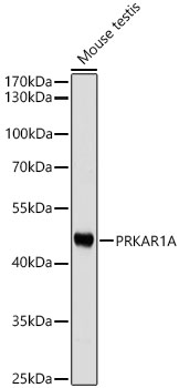 | Western blot analysis of lysates from Mouse testis using PRKAR1A Rabbit pAb (A0906) at 1:1000 dilution. Secondary antibody: HRP-conjugated Goat anti-Rabbit IgG (H+L) (AS014) at 1:10000 dilution. Lysates/proteins: 25 μg per lane. Blocking buffer: 3% nonfat dry milk in TBST. Detection: ECL Basic Kit (RM00020). Exposure time: 10s. |
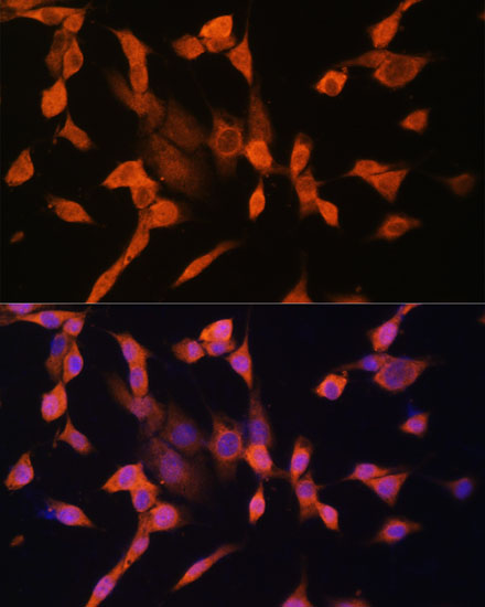 | Immunofluorescence analysis of NIH/3T3 cells using PRKAR1A Rabbit pAb (A0906) at dilution of 1:100. Secondary antibody: Cy3-conjugated Goat anti-Rabbit IgG (H+L) (AS007) at 1:500 dilution. Blue: DAPI for nuclear staining. |
You may also be interested in:

