Your shopping cart is empty!
")
| Reactivity: | Human, Mouse, Rat |
| Applications: | WB, IHC, IF/IC, ELISA |
| Host Species: | Rabbit |
| Isotype: | IgG |
| Clonality: | Polyclonal antibody |
| Gene Name: | polypyrimidine tract binding protein 1 |
| Gene Symbol: | PTBP1 |
| Synonyms: | PTB; PTB2; PTB3; PTB4; pPTB; HNRPI; PTB-1; PTB-T; HNRNPI; HNRNP-I; P1 |
| Gene ID: | 5725 |
| UniProt ID: | P26599 |
| Immunogen: | Recombinant fusion protein containing a sequence corresponding to amino acids 1-290 of human PTBP1 (NP_114368.1). |
| Dilution: | WB 1:500-1:1000; IHC 1:50-1:100; IF/IC 1:50-1:100 |
| Purification Method: | Affinity purification |
| Concentration: | 5.31 mg/ml |
| Buffer: | PBS with 0.02% sodium azide, 50% glycerol ,pH7.3. |
| Storage: | Store at -20°C. Avoid freeze / thaw cycles. |
| Documents: | Manual-PTBP1 polyclonal antibody |
Background
This gene belongs to the subfamily of ubiquitously expressed heterogeneous nuclear ribonucleoproteins (hnRNPs). The hnRNPs are RNA-binding proteins and they complex with heterogeneous nuclear RNA (hnRNA). These proteins are associated with pre-mRNAs in the nucleus and appear to influence pre-mRNA processing and other aspects of mRNA metabolism and transport. While all of the hnRNPs are present in the nucleus, some seem to shuttle between the nucleus and the cytoplasm. The hnRNP proteins have distinct nucleic acid binding properties. The protein encoded by this gene has four repeats of quasi-RNA recognition motif (RRM) domains that bind RNAs. This protein binds to the intronic polypyrimidine tracts that requires pre-mRNA splicing and acts via the protein degradation ubiquitin-proteasome pathway. It may also promote the binding of U2 snRNP to pre-mRNAs. This protein is localized in the nucleoplasm and it is also detected in the perinucleolar structure. Alternatively spliced transcript variants encoding different isoforms have been described.
Images
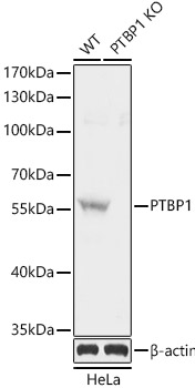
 | Western blot analysis of lysates from wild type (WT) and PTBP1 knockout (KO) HeLa cells, using [KO Validated] PTBP1 Rabbit pAb (A1831) at 1:1000 dilution. Secondary antibody: HRP-conjugated Goat anti-Rabbit IgG (H+L) (AS014) at 1:10000 dilution. Lysates/proteins: 25μg per lane. Blocking buffer: 3% nonfat dry milk in TBST. Detection: ECL Basic Kit (RM00020). Exposure time: 5s. |
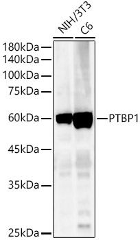
 | Western blot analysis of various lysates, using [KO Validated] PTBP1 Rabbit pAb (A1831) at 1:400 dilution. Secondary antibody: HRP-conjugated Goat anti-Rabbit IgG (H+L) (AS014) at 1:10000 dilution. Lysates/proteins: 25μg per lane. Blocking buffer: 3% nonfat dry milk in TBST. Detection: ECL Basic Kit (RM00020). Exposure time: 180s. |
 | Western blot analysis of lysates from wild type(WT) and PTBP1 knockout (KO) HeLa(KO) cells, using [KO Validated] PTBP1 Rabbit pAb (A1831) at 1:1000 dilution. Secondary antibody: HRP-conjugated Goat anti-Rabbit IgG (H+L) (AS014) at 1:10000 dilution. Lysates/proteins: 25μg per lane. Blocking buffer: 3% nonfat dry milk in TBST. Detection: ECL Basic Kit (RM00020). Exposure time: 1s. |
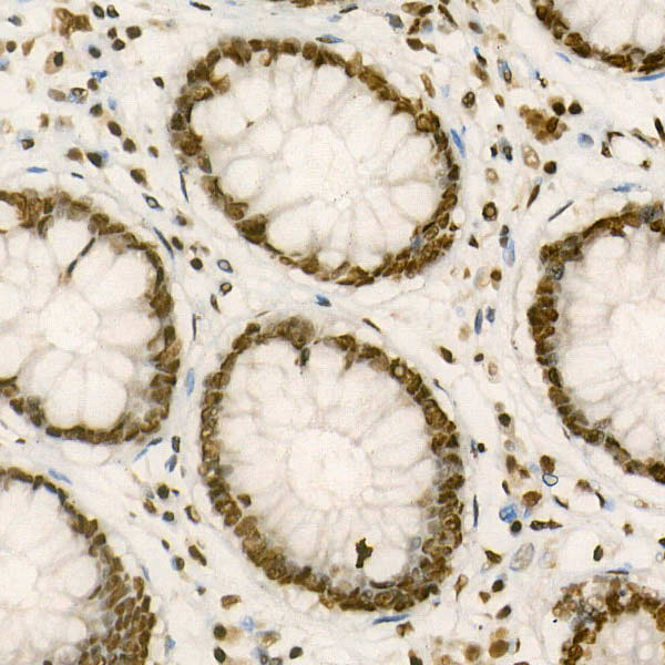
 | Immunohistochemistry analysis of paraffin-embedded Human colon carcinoma using [KO Validated] PTBP1 Rabbit pAb (A1831) at dilution of 1:25 (40x lens). High pressure antigen retrieval performed with 0.01M Citrate Bufferr (pH 6.0) prior to IHC staining. |
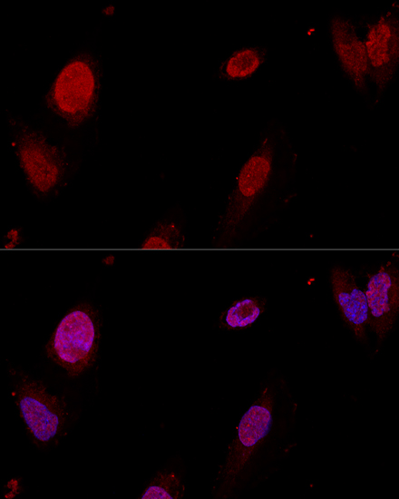
 | Confocal immunofluorescence analysis of U-2OS cells using [KO Validated] PTBP1 Rabbit pAb (A1831) at dilution of 1:50. Blue: DAPI for nuclear staining. |
You may also be interested in:

