Your shopping cart is empty!
| Reactivity: | Human |
| Applications: | WB, ELISA |
| Host Species: | Rabbit |
| Isotype: | IgG |
| Clonality: | Monoclonal antibody |
| Gene Name: | RAB12, member RAS oncogene family |
| Gene Symbol: | RAB12 |
| Synonyms: | RAB12 |
| Gene ID: | 201475 |
| UniProt ID: | Q6IQ22 |
| Clone ID: | 2R5P3 |
| Immunogen: | Recombinant protein containing a sequence corresponding to amino acids 1-244 of human Rab12 protein (Q6IQ22). |
| Purification Method: | Affinity purification |
| Concentration: | 1.09 mg/ml |
| Buffer: | PBS with 0.09% Sodium azide, 0.05% BSA, 50% glycerol, pH7.3 |
| Storage: | Store at -20°C. Avoid freeze / thaw cycles. |
| Documents: | Manual-RAB12 monoclonal antibody |
Background
Rab12 is a member of the RAS oncogene family and plays a crucial role in intracellular membrane trafficking. This small GTPase protein is involved in the transport of vesicles, particularly from recycling endosomes to lysosomes, and is essential for processes like autophagy and the degradation of the transferrin receptor. Rab12 cycles between an inactive GDP-bound form and an active GTP-bound form, recruiting various effectors responsible for vesicle formation, movement, and fusion. In addition to being a substrate for phosphorylation by LRRK2 (Leucine-Rich Repeat Kinase 2), Rab12 has recently been identified as its significant regulator. It has been shown that Rab12 can activate LRRK2, leading to increased phosphorylation of Rab10, another protein involved in intracellular trafficking. This activation is particularly notable in the context of lysosomal damage, where Rab12 helps recruit LRRK2 to lysosomes, enhancing its activity. Dysregulation of this pathway due to pathogenic LRRK2 mutations can contribute to the neurodegenerative processes observed in Parkinson's disease.
Images
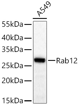 | Western blot analysis of lysates from A549 cells using Rab12 Rabbit mAb (A26172) at 1:2000 dilution incubated at room temperature for 1.5 hours. Secondary antibody: HRP-conjugated Goat anti-Rabbit IgG (H+L) (AS014) at 1:10000 dilution. Lysates/proteins: 25 μg per lane. Blocking buffer: 3% nonfat dry milk in TBST. Detection: ECL Basic Kit (RM00020). Exposure time: 60s. |
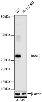 | Western blot analysis of lysates from wild type (WT) and Rab12 knockout (KO) A-549 cells using Rab12 Rabbit mAb (A26172) at 1:100000 dilution incubated overnight at 4℃. Secondary antibody: IRDye® 800CW Goat anti-Rabbit IgG Secondary Antibody (926-32211) at 1:25000 dilution. Lysates/proteins: 15 μg per lane. Blocking buffer: 5% nonfat dry milk in TBST. Detection: LI-COR Odyssey CLx. Exposure time: LI-COR scan using automatic sensitivity setting. |
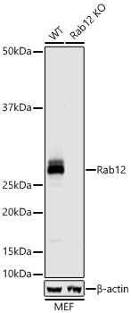 | Western blot analysis of lysates from wild type (WT) and Rab12 knockout (KO) MEF cells using Rab12 Rabbit mAb (A26172) at 1:100000 dilution incubated overnight at 4℃ Secondary antibody: IRDye® 800CW Goat anti-Rabbit IgG Secondary Antibody (926-32211) at 1:25000 dilution. Lysates/proteins: 15 μg per lane. Blocking buffer: 5% nonfat dry milk in TBST. Detection: LI-COR Odyssey CLx. Exposure time: LI-COR scan using automatic sensitivity setting. |
You may also be interested in:

