Your shopping cart is empty!
")
KO-Validated NF-kB p65/RelA Rabbit mAb (20 μl)
| Reactivity: | Human, Mouse, Rat, Monkey |
| Applications: | WB, IHC, IF/IC, ELISA, ChIP |
| Host Species: | Rabbit |
| Isotype: | IgG |
| Clonality: | Monoclonal antibody |
| Gene Name: | RELA proto-oncogene, NF-kB subunit |
| Gene Symbol: | RELA |
| Synonyms: | p65; CMCU; NFKB3; AIF3BL3; NF-kB p65/RelA |
| Gene ID: | 5970 |
| UniProt ID: | Q04206 |
| Clone ID: | 5P4U2 |
| Immunogen: | A synthetic peptide corresponding to a sequence within amino acids 450-551 of human NF-κB p65 (Q04206). |
| Dilution: | WB 1:5000-1:20000; IHC 1:500-1:2000; IF/IC 1:50-1:200 |
| Purification Method: | Affinity purification |
| Concentration: | 1.00 mg/mL |
| Buffer: | PBS with 0.09% sodium azid, 0.05% BSA, 50% glycerol, pH7.3. |
| Storage: | Store at -20°C. Avoid freeze / thaw cycles. |
| Documents: | Manual-RELA monoclonal antibody |
Background
NF-kappa-B is a ubiquitous transcription factor involved in several biological processes. It is held in the cytoplasm in an inactive state by specific inhibitors. Upon degradation of the inhibitor, NF-kappa-B moves to the nucleus and activates transcription of specific genes. NF-kappa-B is composed of NFKB1 or NFKB2 bound to either REL, RELA, or RELB. The most abundant form of NF-kappa-B is NFKB1 complexed with the product of this gene, RELA. Four transcript variants encoding different isoforms have been found for this gene.
Images
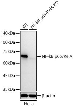 | Western blot analysis of lysates from wild type (WT) and NF-kB p65/RelA knockout (KO) HeLa cells using NF-kB p65/RelA Rabbit mAb (A19653) at 1:10000 dilution incubated overnight at 4℃. Secondary antibody: HRP-conjugated Goat anti-Rabbit IgG (H+L) (AS014) at 1:10000 dilution. Lysates/proteins: 25 μg per lane. Blocking buffer: 3% nonfat dry milk in TBST. Detection: ECL Basic Kit (RM00020). Exposure time: 30s. |
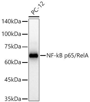 | Western blot analysis of lysates from PC-12 cells using NF-kB p65/RelA Rabbit mAb (A19653) at 1:10000 dilution incubated overnight at 4℃. Secondary antibody: HRP-conjugated Goat anti-Rabbit IgG (H+L) (AS014) at 1:10000 dilution. Lysates/proteins: 25 μg per lane. Blocking buffer: 3% nonfat dry milk in TBST. Detection: ECL Basic Kit (RM00020). Exposure time: 30s. |
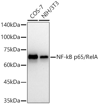 | Western blot analysis of various lysates using NF-kB p65/RelA Rabbit mAb (A19653) at 1:10000 dilution incubated overnight at 4℃. Secondary antibody: HRP-conjugated Goat anti-Rabbit IgG (H+L) (AS014) at 1:10000 dilution. Lysates/proteins: 25 μg per lane. Blocking buffer: 3% nonfat dry milk in TBST. Detection: ECL Basic Kit (RM00020). Exposure time: 60s. |
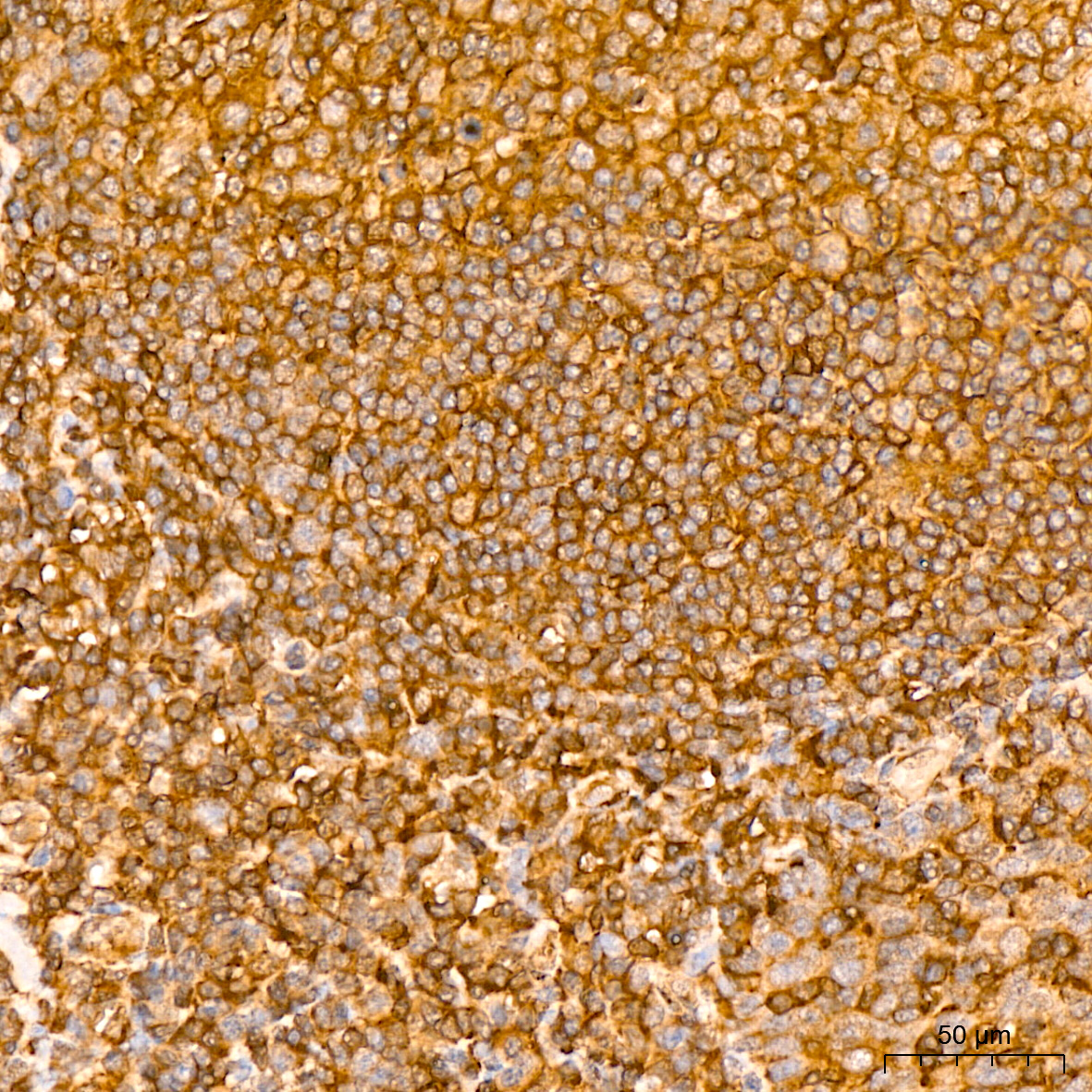
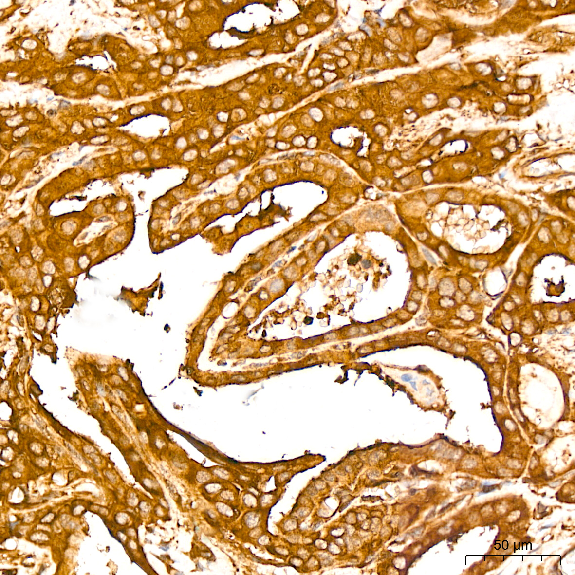 | Immunohistochemistry analysis of paraffin-embedded Human thyroid cancer tissue using NF-kB p65/RelA Rabbit mAb (A19653) at a dilution of 1:800 (40x lens). High pressure antigen retrieval performed with 0.01M Citrate Bufferr (pH 6.0) prior to IHC staining. |
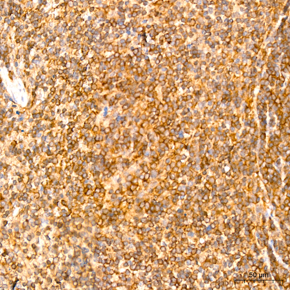
 | Immunohistochemistry analysis of paraffin-embedded Human tonsil tissue using NF-kB p65/RelA Rabbit mAb (A19653) at a dilution of 1:800 (40x lens). High pressure antigen retrieval performed with 0.01M Citrate Bufferr (pH 6.0) prior to IHC staining. |
 | Immunohistochemistry analysis of paraffin-embedded Mouse spleen tissue using NF-kB p65/RelA Rabbit mAb (A19653) at a dilution of 1:800 (40x lens). High pressure antigen retrieval performed with 0.01M Citrate Bufferr (pH 6.0) prior to IHC staining. |
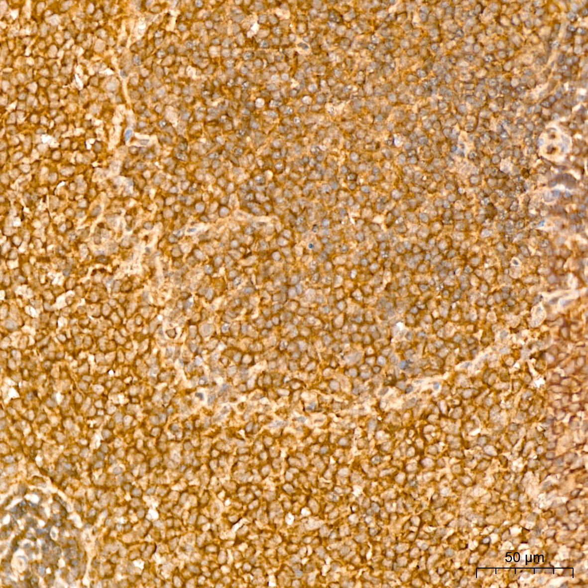 | Immunohistochemistry analysis of paraffin-embedded Rat spleen tissue using NF-kB p65/RelA Rabbit mAb (A19653) at a dilution of 1:800 (40x lens). High pressure antigen retrieval performed with 0.01M Citrate Bufferr (pH 6.0) prior to IHC staining. |
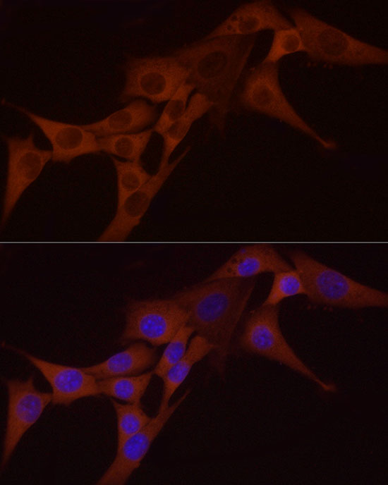 | Immunofluorescence analysis of NIH/3T3 cells using NF-kB p65/RelA Rabbit mAb (A19653) at dilution of 1:100 (40x lens). Secondary antibody: Cy3-conjugated Goat anti-Rabbit IgG (H+L) (AS007) at 1:500 dilution. Blue: DAPI for nuclear staining. |
You may also be interested in:

