Your shopping cart is empty!
")
| Reactivity: | Human, Mouse, Rat |
| Applications: | WB, IHC, IP, ELISA, ChIP, CUT&Tag |
| Host Species: | Rabbit |
| Isotype: | IgG |
| Clonality: | Monoclonal antibody |
| Gene Name: | SMAD family member 4 |
| Gene Symbol: | SMAD4 |
| Synonyms: | JIP; DPC4; MADH4; MYHRS; d4 |
| Gene ID: | 4089 |
| UniProt ID: | Q13485 |
| Clone ID: | 6B4D6 |
| Immunogen: | Recombinant fusion protein containing a sequence corresponding to amino acids 320-552 of human Smad4 (NP_005350.1). |
| Dilution: | WB 1:1000-1:6000; IHC 1:500-1:2000 |
| Purification Method: | Affinity purification |
| Concentration: | 1 mg/ml |
| Buffer: | PBS with 0.05% proclin300, 0.05% BSA, 50% glycerol, pH7.3. |
| Storage: | Store at -20°C. Avoid freeze / thaw cycles. |
| Documents: | Manual-SMAD4 monoclonal antibody |
Background
This gene encodes a member of the Smad family of signal transduction proteins. Smad proteins are phosphorylated and activated by transmembrane serine-threonine receptor kinases in response to transforming growth factor (TGF)-beta signaling. The product of this gene forms homomeric complexes and heteromeric complexes with other activated Smad proteins, which then accumulate in the nucleus and regulate the transcription of target genes. This protein binds to DNA and recognizes an 8-bp palindromic sequence (GTCTAGAC) called the Smad-binding element (SBE). The protein acts as a tumor suppressor and inhibits epithelial cell proliferation. It may also have an inhibitory effect on tumors by reducing angiogenesis and increasing blood vessel hyperpermeability. The encoded protein is a crucial component of the bone morphogenetic protein signaling pathway. The Smad proteins are subject to complex regulation by post-translational modifications. Mutations or deletions in this gene have been shown to result in pancreatic cancer, juvenile polyposis syndrome, and hereditary hemorrhagic telangiectasia syndrome.
Images
 | CUT&Tag was performed using the CUT&Tag Assay Kit (pAG-Tn5) for illumina (RK20265) from 10⁵ K562 cells with 1ug μg of [KO Validated] Smad4 Rabbit mAb (A19116), followed by incubation with Goat Anti-Rabbit IgG(H+L)(AS070). The CUT&Tag results denote the enrichment pattern of [KO Validated] Smad4 Rabbit mAb across chromosome 19 (upper panel) and the genomic region encompassing JUNB, a representative gene enriched in [KO Validated] Smad4 Rabbit mAb (lower panel). |
 | CUT&Tag was performed using the CUT&Tag Assay Kit (pAG-Tn5) for Illumina (RK20265) from10⁵ K562 with 1 μg of [KO Validated] Smad4 Rabbit mAb (A19116), followed by incubation with Goat Anti-Rabbit IgG(H+L)(AS070).The results denote the enrichment pattern of Smad4 around JUNB gene. |
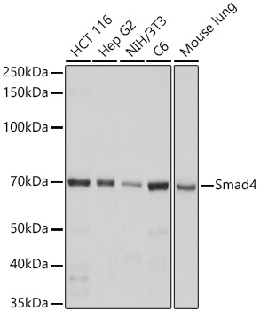 | Western blot analysis of various lysates using [KO Validated] Smad4 Rabbit mAb (A19116) at 1:1000 dilution incubated overnight at 4℃. Secondary antibody: HRP-conjugated Goat anti-Rabbit IgG (H+L) (AS014) at 1:10000 dilution. Lysates/proteins: 25 μg per lane. Blocking buffer: 3% nonfat dry milk in TBST. Detection: ECL Basic Kit (RM00020). Exposure time: 3s. |
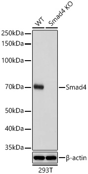 | Western blot analysis of lysates from wild type (WT) and Smad4 knockout (KO) 293T cells using [KO Validated] Smad4 Rabbit mAb (A19116) at 1:1000 dilution incubated overnight at 4℃. Secondary antibody: HRP-conjugated Goat anti-Rabbit IgG (H+L) (AS014) at 1:10000 dilution. Lysates/proteins: 25 μg per lane. Blocking buffer: 3% nonfat dry milk in TBST. Detection: ECL Basic Kit (RM00020). Exposure time: 3s. |
 | Immunohistochemistry analysis of paraffin-embedded Human placenta tissue using [KO Validated] Smad4 Rabbit mAb (A19116) at a dilution of 1:500 (40x lens). High pressure antigen retrieval performed with 0.01M Tris-EDTA Buffer (pH 9.0) prior to IHC staining. |
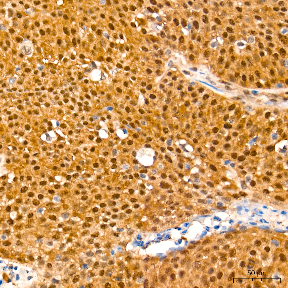 | Immunohistochemistry analysis of paraffin-embedded Human cervix cancer tissue using [KO Validated] Smad4 Rabbit mAb (A19116) at a dilution of 1:500 (40x lens). High pressure antigen retrieval performed with 0.01M Tris-EDTA Buffer (pH 9.0) prior to IHC staining. |
 | Immunohistochemistry analysis of paraffin-embedded Mouse testis tissue using [KO Validated] Smad4 Rabbit mAb (A19116) at a dilution of 1:500 (40x lens). High pressure antigen retrieval performed with 0.01M Tris-EDTA Buffer (pH 9.0) prior to IHC staining. |
 | Immunohistochemistry analysis of paraffin-embedded Rat testis tissue using [KO Validated] Smad4 Rabbit mAb (A19116) at a dilution of 1:500 (40x lens). High pressure antigen retrieval performed with 0.01M Tris-EDTA Buffer (pH 9.0) prior to IHC staining. |
You may also be interested in:

