Your shopping cart is empty!
")
KO-Validated SMARCB1/SNF5 Rabbit mAb (20 μl)
| Reactivity: | Human, Mouse, Rat |
| Applications: | WB, IHC, IP, ELISA |
| Host Species: | Rabbit |
| Isotype: | IgG |
| Clonality: | Monoclonal antibody |
| Gene Name: | SWI/SNF related BAF chromatin remodeling complex subunit B1 |
| Gene Symbol: | SMARCB1 |
| Synonyms: | RDT; CSS3; INI1; SNF5; Snr1; BAF47; INI-1; MRD15; RTPS1; Sfh1p; hSNFS; SNF5L1; SWNTS1; PPP1R144; F5 |
| Gene ID: | 6598 |
| UniProt ID: | Q12824 |
| Clone ID: | 9A9G0 |
| Immunogen: | A synthetic peptide corresponding to a sequence within amino acids 50-150 of human SMARCB1/SNF5 (NP_003064.2). |
| Dilution: | WB 1:1000-1:5000; IHC 1:50-1:200 |
| Purification Method: | Affinity purification |
| Concentration: | 1.20 mg/mL |
| Buffer: | PBS with 0.05% proclin300, 0.05% BSA, 50% glycerol, pH7.3. |
| Storage: | Store at -20°C. Avoid freeze / thaw cycles. |
| Documents: | Manual-SMARCB1 monoclonal antibody |
Background
The protein encoded by this gene is part of a complex that relieves repressive chromatin structures, allowing the transcriptional machinery to access its targets more effectively. The encoded nuclear protein may also bind to and enhance the DNA joining activity of HIV-1 integrase. This gene has been found to be a tumor suppressor, and mutations in it have been associated with malignant rhabdoid tumors. Alternatively spliced transcript variants have been found for this gene.
Images
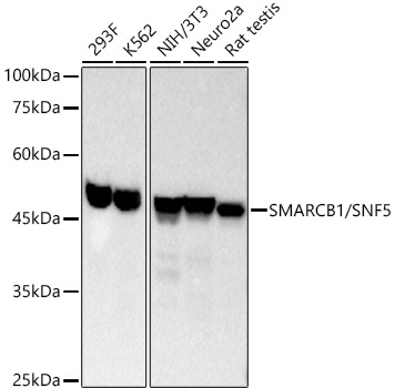 | Western blot analysis of various lysates, using SMARCB1/SNF5 Rabbit mAb (A3247) at 1:2000 dilution. Secondary antibody: HRP-conjugated Goat anti-Rabbit IgG (H+L) (AS014) at 1:2000 dilution. Lysates/proteins: 25μg per lane. Blocking buffer: 3% nonfat dry milk in TBST. Detection: ECL Basic Kit (RM00020). Exposure time: 10s. |
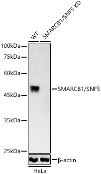 | Western blot analysis of lysates from wild type(WT) and SMARCB1/SNF5 Rabbit mAb knockout (KO) HeLa(KO) cells, using SMARCB1/SNF5 Rabbit mAb (A3247) at 1:2000 dilution. Secondary antibody: HRP-conjugated Goat anti-Rabbit IgG (H+L) (AS014) at 1:10000 dilution. Lysates/proteins: 25μg per lane. Blocking buffer: 3% nonfat dry milk in TBST. Detection: ECL Basic Kit (RM00020). Exposure time: 10s. |
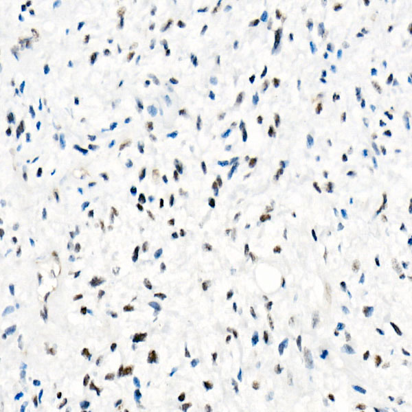 | Immunohistochemistry analysis of paraffin-embedded Human epithelioid sarcoma (ini-1 deletion) using [KO Validated] SMARCB1/SNF5 Rabbit mAb (A3247) at dilution of 1:100 (40x lens). High pressure antigen retrieval performed with 0.01M Tris/EDTA Buffer (pH 9.0) prior to IHC staining. |
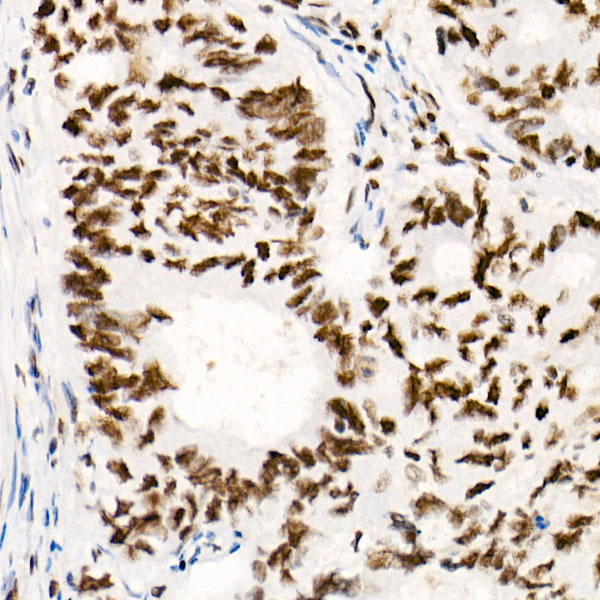 | Immunohistochemistry analysis of paraffin-embedded Human colon carcinoma using [KO Validated] SMARCB1/SNF5 Rabbit mAb (A3247) at dilution of 1:100 (40x lens). High pressure antigen retrieval performed with 0.01M Tris/EDTA Buffer (pH 9.0) prior to IHC staining. |
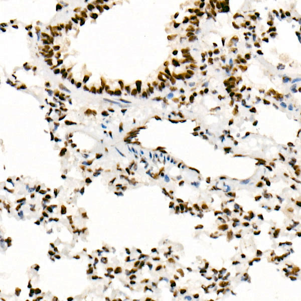 | Immunohistochemistry analysis of paraffin-embedded Rat lung using [KO Validated] SMARCB1/SNF5 Rabbit mAb (A3247) at dilution of 1:100 (40x lens). High pressure antigen retrieval performed with 0.01M Tris/EDTA Buffer (pH 9.0) prior to IHC staining. |
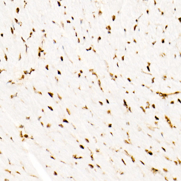 | Immunohistochemistry analysis of paraffin-embedded Mouse heart using [KO Validated] SMARCB1/SNF5 Rabbit mAb (A3247) at dilution of 1:100 (40x lens). High pressure antigen retrieval performed with 0.01M Tris/EDTA Buffer (pH 9.0) prior to IHC staining. |
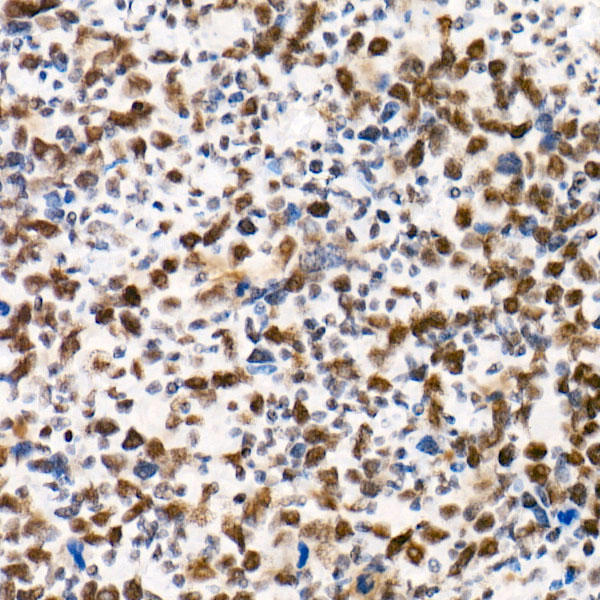 | Immunohistochemistry analysis of paraffin-embedded Human undifferentiated carcinoma of esophagus using [KO Validated] SMARCB1/SNF5 Rabbit mAb (A3247) at dilution of 1:100 (40x lens). High pressure antigen retrieval performed with 0.01M Tris/EDTA Buffer (pH 9.0) prior to IHC staining. |
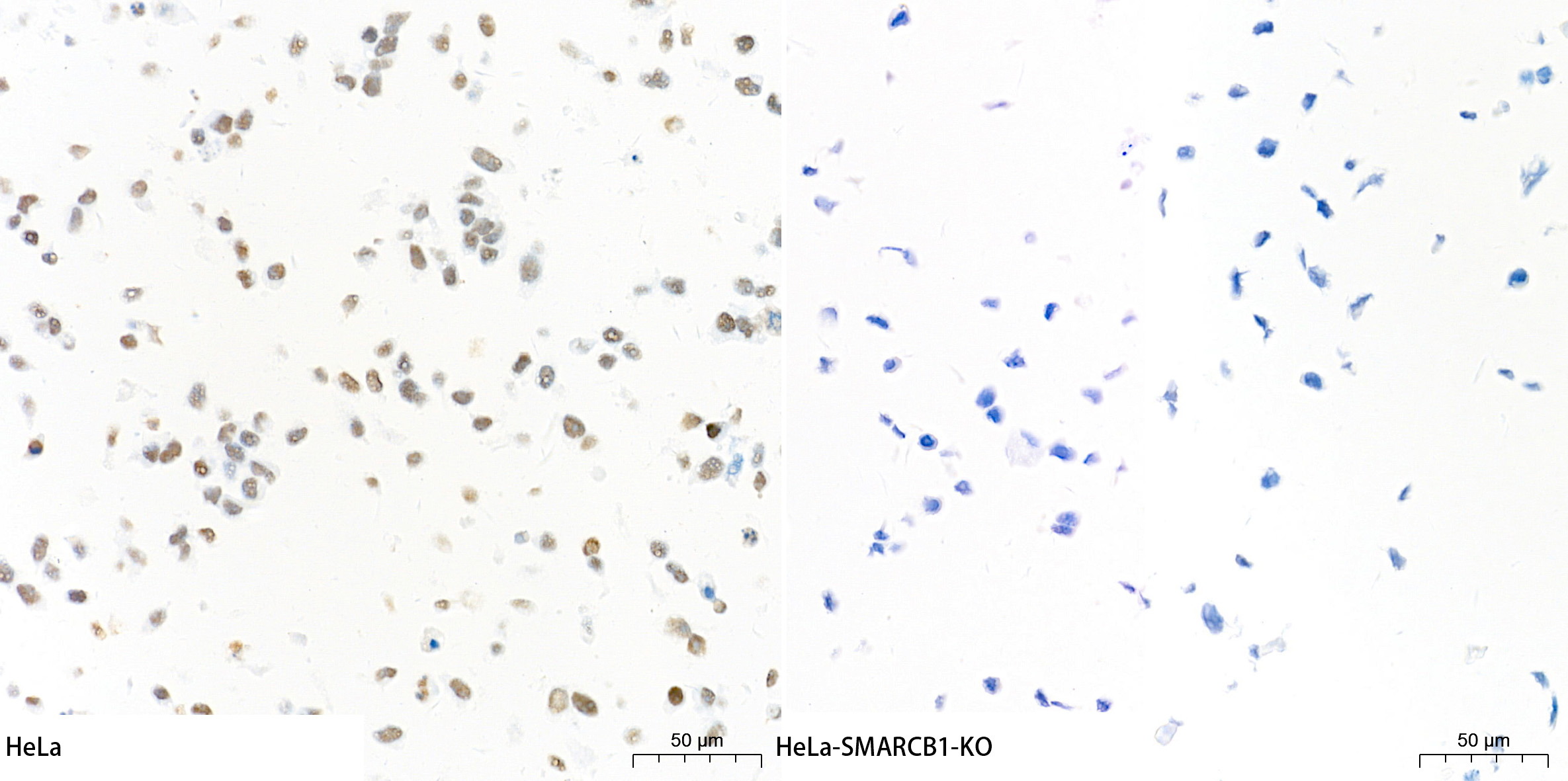 | Immunohistochemistry analysis of paraffin-embedded HeLa and HeLa-SMARCB1-KO cells using [KO Validated] SMARCB1/SNF5 Rabbit mAb (A3247) at a dilution of 1:1000 (40x lens). High pressure antigen retrieval performed with 0.01M Tris-EDTA Buffer (pH 9.0) prior to IHC staining. |
You may also be interested in:

